Mesa Kelly J, Selmic Laura E, Pande Paritosh, Monroy Guillermo L, Reagan Jennifer, Samuelson Jonathan, Driskell Elizabeth, Li Joanne, Marjanovic Marina, Chaney Eric J, Boppart Stephen A
Beckman Institute for Advanced Science and Technology, University of Illinois at Urbana-Champaign, Urbana, Illinois.
Department of Electrical and Computer Engineering, University of Illinois at Urbana-Champaign, Urbana, Illinois.
Lasers Surg Med. 2017 Mar;49(3):240-248. doi: 10.1002/lsm.22633. Epub 2017 Mar 20.
Sarcomas are rare but highly aggressive tumors, and local recurrence after surgical excision can occur in up to 50% cases. Therefore, there is a strong clinical need for accurate tissue differentiation and margin assessment to reduce incomplete resection and local recurrence. The purpose of this study was to investigate the use of optical coherence tomography (OCT) and a novel image texture-based processing algorithm to differentiate sarcoma from muscle and adipose tissue.
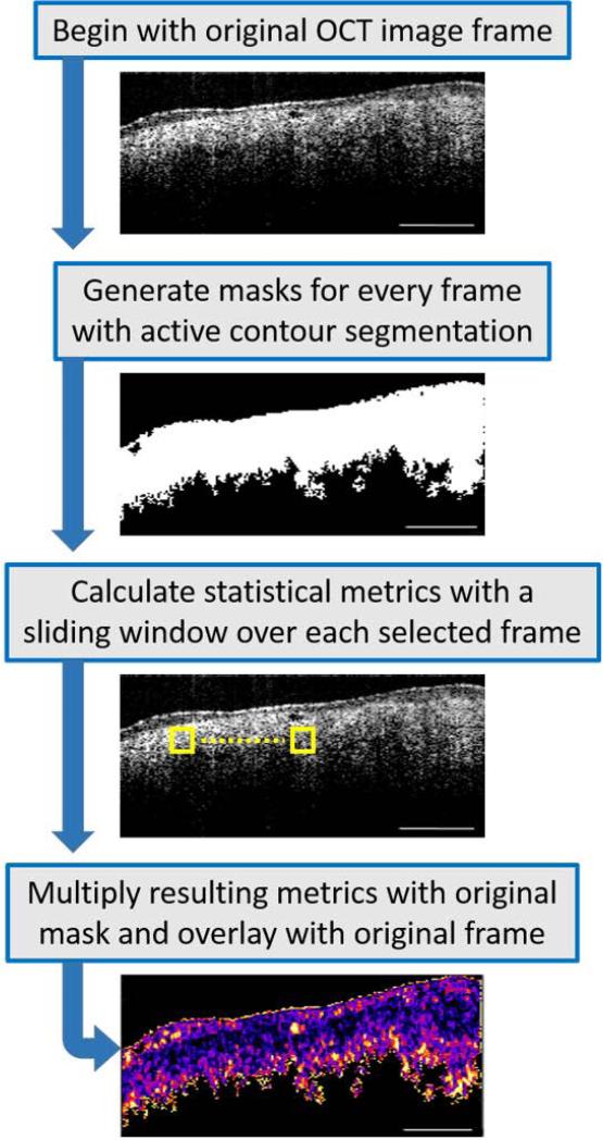
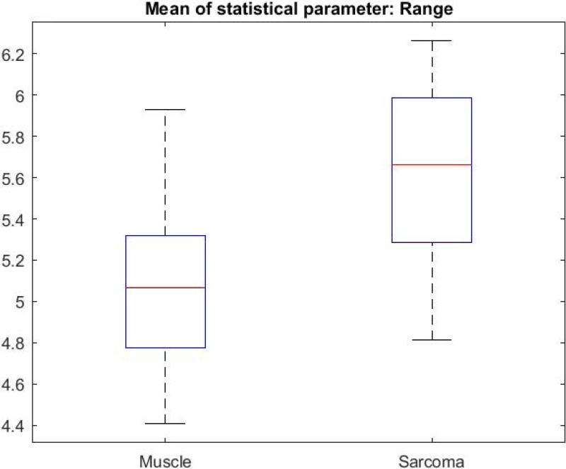
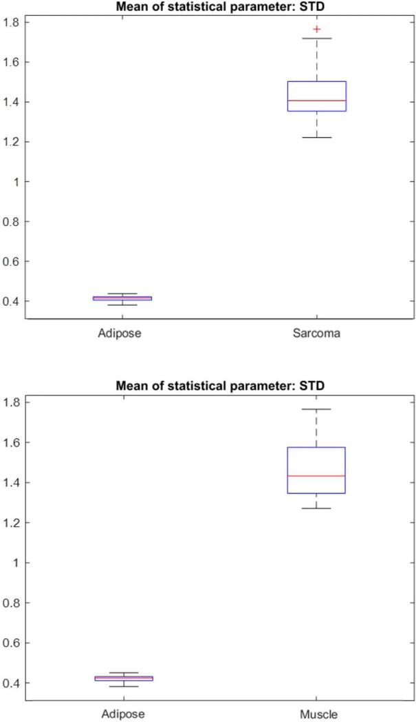
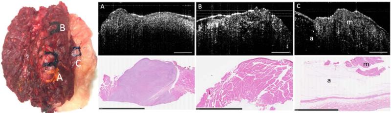
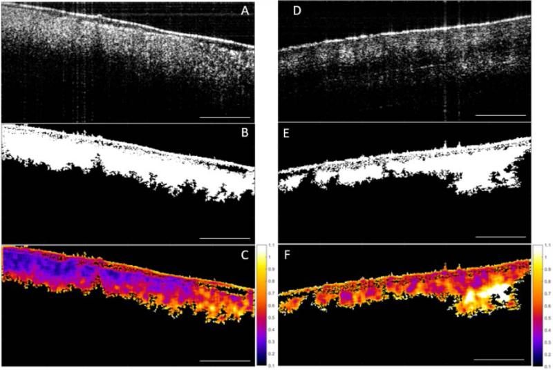
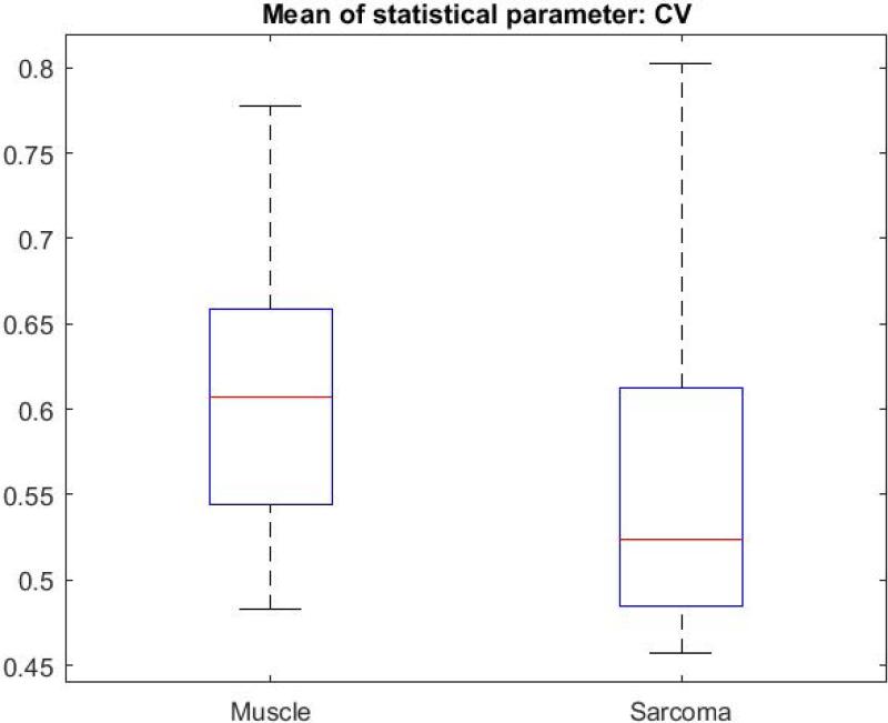
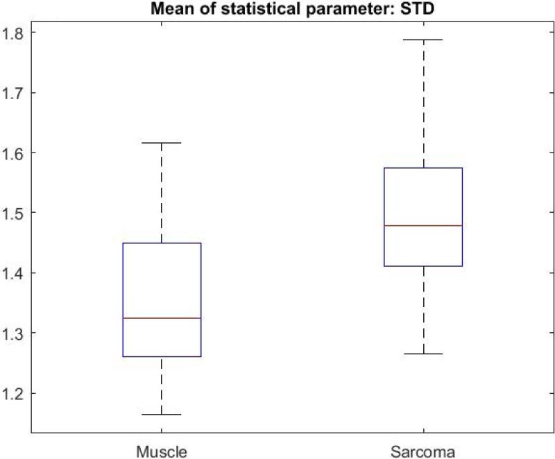
In this study, tumor margin delineation in 19 feline and canine veterinary patients was achieved with intraoperative OCT to help validate tumor resection. While differentiation of lower-scattering adipose tissue from higher-scattering muscle and tumor tissue was relatively straightforward, it was more challenging to distinguish between dense highly scattering muscle and tumor tissue types based on scattering intensity and microstructural features alone. To improve tissue-type differentiation in a more objective and automated manner, three descriptive statistical metrics, namely the coefficient of variation (CV), standard deviation (STD), and Range, were implemented in a custom algorithm applied to the OCT images.
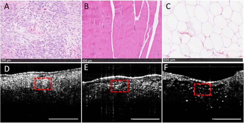
Over 22,800 OCT images were collected intraoperatively from over 38 sites on 19 ex vivo tissue specimens removed during sarcoma surgeries. Following the generation of an initial set of OCT images correlated with standard hematoxylin and eosin-stained histopathology, over 760 images were subsequently used for automated analysis. Using texture-based image processing metrics, OCT images of sarcoma, muscle, and adipose tissue were all found to be statistically different from one another (P ≤ 0.001).
These results demonstrate the potential of using intraoperative OCT, along with an automated tissue differentiation algorithm, as a guidance tool for soft tissue sarcoma margin delineation in the operating room. Lasers Surg. Med. 49:240-248, 2017. © 2017 Wiley Periodicals, Inc.
肉瘤虽罕见但侵袭性强,手术切除后局部复发率可达50%。因此,临床上迫切需要准确的组织分化和切缘评估,以减少切除不彻底和局部复发。本研究旨在探讨光学相干断层扫描(OCT)及一种基于图像纹理的新型处理算法在肉瘤与肌肉及脂肪组织鉴别中的应用。
本研究中,对19例猫科和犬科兽医患者进行术中OCT以实现肿瘤切缘勾画,从而辅助验证肿瘤切除情况。区分低散射的脂肪组织与高散射的肌肉和肿瘤组织相对简单,但仅基于散射强度和微观结构特征来区分致密的高散射肌肉和肿瘤组织类型则更具挑战性。为以更客观和自动化的方式改善组织类型鉴别,在应用于OCT图像的自定义算法中采用了三个描述性统计指标,即变异系数(CV)、标准差(STD)和极差。
术中从肉瘤手术切除的19个离体组织标本的38个以上部位采集了超过22,800张OCT图像。在生成与标准苏木精和伊红染色组织病理学相关的初始OCT图像集后,随后对超过760张图像进行了自动分析。使用基于纹理的图像处理指标发现,肉瘤、肌肉和脂肪组织的OCT图像在统计学上均彼此不同(P≤0.001)。
这些结果表明,术中OCT与自动组织分化算法相结合,有潜力作为手术室软组织肉瘤切缘勾画的指导工具。《激光外科与医学》2017年第49卷:240 - 248页。©2017威利期刊公司。