Center for Medical Image Science and Visualization (CMIV), Linköping University, Linköpings universitet/US, SE-581 85, Linköping, SE, Sweden.
Department of Medical and Health Sciences (IMH), Linköping University, Linköping, Sweden.
Brain Imaging Behav. 2018 Apr;12(2):411-424. doi: 10.1007/s11682-017-9706-y.
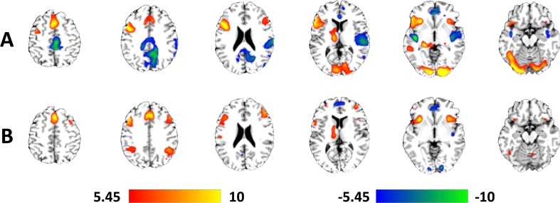
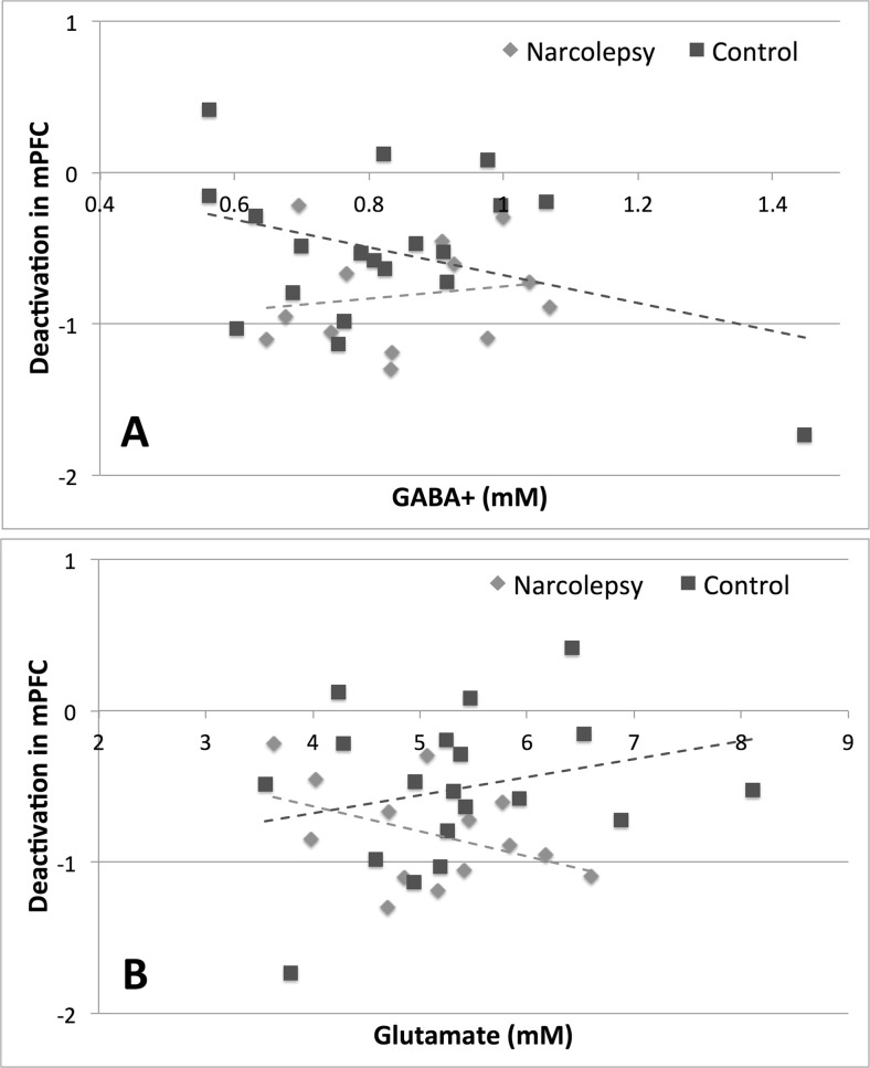
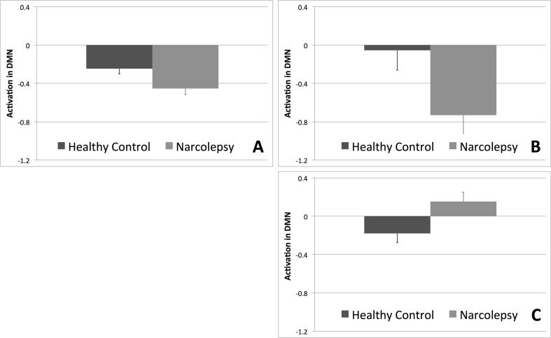
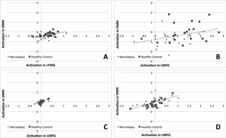
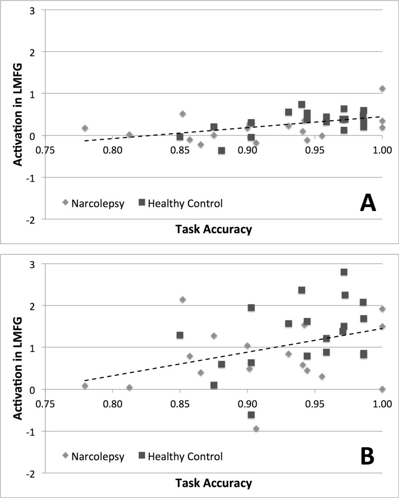
The study investigated brain activity changes during performance of a verbal working memory task in a population of adolescents with narcolepsy. Seventeen narcolepsy patients and twenty healthy controls performed a verbal working memory task during simultaneous fMRI and EEG acquisition. All subjects also underwent MRS to measure GABA and Glutamate concentrations in the medial prefrontal cortex. Activation levels in the default mode network and left middle frontal gyrus were examined to investigate whether narcolepsy is characterized by an imbalance in cognitive resources. Significantly increased deactivation within the default mode network during task performance was observed for the narcolepsy patients for both the encoding and recognition phases of the task. No evidence for task performance deficits or reduced activation within the left middle frontal gyrus was noted for the narcolepsy patients. Correlation analyses between the spectroscopy and fMRI data indicated that deactivation of the anterior aspect of the default mode in narcolepsy patients correlated more with increased concentrations of Glutamate and decreased concentrations of GABA. In contrast, deactivation in the default mode was correlated with increased concentrations of GABA and decreased concentrations of Glutamate in controls. The results suggested that narcolepsy is not characterized by a deficit in working memory but rather an imbalance of cognitive resources in favor of monitoring and maintaining attention over actual task performance. This points towards dysregulation within the sustained attention system being the origin behind self-reported cognitive difficulties in narcolepsy.
该研究调查了嗜睡症青少年群体在执行口头工作记忆任务时大脑活动的变化。17 名嗜睡症患者和 20 名健康对照者在同时进行 fMRI 和 EEG 采集时执行了口头工作记忆任务。所有受试者还接受了 MRS 以测量内侧前额叶皮质中的 GABA 和谷氨酸浓度。研究了默认模式网络和左额中回的激活水平,以调查嗜睡症是否表现出认知资源失衡。观察到嗜睡症患者在任务执行期间默认模式网络内的去激活显著增加,无论是在任务的编码还是识别阶段。未发现嗜睡症患者在任务表现上存在缺陷或左额中回的激活减少。频谱和 fMRI 数据之间的相关分析表明,嗜睡症患者默认模式前部的去激活与谷氨酸浓度增加和 GABA 浓度降低更相关。相比之下,在对照组中,默认模式的去激活与 GABA 浓度增加和谷氨酸浓度降低相关。研究结果表明,嗜睡症不是工作记忆缺陷,而是认知资源失衡,有利于监控和维持注意力而不是实际的任务表现。这表明,维持注意力系统的失调是嗜睡症患者自我报告认知困难的根源。