Lohrke Jessica, Frisk Anna-Lena, Frenzel Thomas, Schöckel Laura, Rosenbruch Martin, Jost Gregor, Lenhard Diana Constanze, Sieber Martin A, Nischwitz Volker, Küppers Astrid, Pietsch Hubertus
From the *MR and CT Contrast Media Research, †Research Pathology Berlin, and ‡Medical and Clinical Affairs Radiology, Bayer AG, Berlin; §Special Histopathology, Bayer AG, Elberfeld; ∥Institute of Vegetative Physiology, Charité; ¶Clinical Project Management, Bayer AG, Berlin; and #Central Institute for Engineering, Electronics and Analytics (ZEA-3), Forschungszentrum Juelich GmbH, Juelich, Germany.
Invest Radiol. 2017 Jun;52(6):324-333. doi: 10.1097/RLI.0000000000000344.
Retrospective studies in patients with primary brain tumors or other central nervous system pathologies as well as postmortem studies have suggested that gadolinium (Gd) deposition occurs in the dentate nucleus (DN) and globus pallidus (GP) after multiple administrations of primarily linear Gd-based contrast agents (GBCAs). However, this deposition has not been associated with any adverse effects or histopathological alterations. The aim of this preclinical study was to systematically examine differences between linear and macrocyclic GBCAs in their potential to induce changes in brain and skin histology including Gd distribution in high spatial resolution.
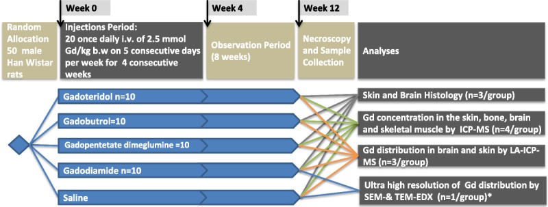
Fifty male Wistar-Han rats were randomly allocated into control (saline, n = 10 rats) and 4 GBCA groups (linear GBCAs: gadodiamide and gadopentetate dimeglumine, macrocyclic GBCAs: gadobutrol and gadoteridol; n = 10 rats per group). The animals received 20 daily intravenous injections at a dose of 2.5 mmol Gd/kg body weight. Eight weeks after the last GBCA administration, the animals were killed, and the brain and skin samples were histopathologically assessed (hematoxylin and eosin; cresyl violet [Nissl]) and by immunohistochemistry. The Gd concentration in the skin, bone, brain, and skeletal muscle samples were analyzed using inductively coupled plasma mass spectroscopy (ICP-MS, n = 4). The spatial Gd distribution in the brain and skin samples was analyzed in cryosections using laser ablation coupled with ICP-MS (LA-ICP-MS, n = 3). For the ultra-high resolution of Gd distribution, brain sections of rats injected with gadodiamide or saline (n = 1) were assessed by scanning electron microscopy coupled to energy dispersive x-ray spectroscopy and transmission electron microscopy, respectively.
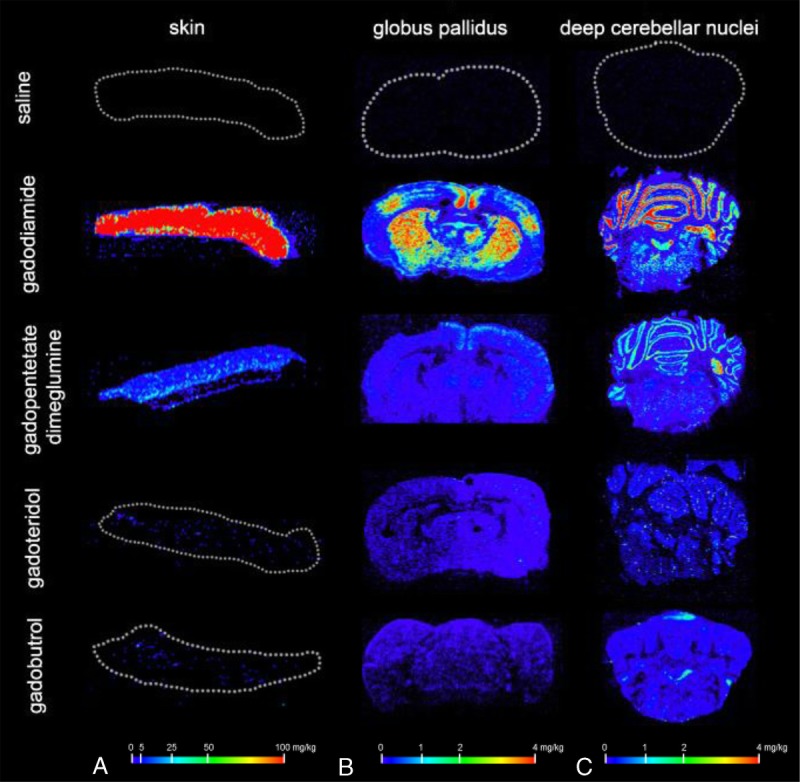
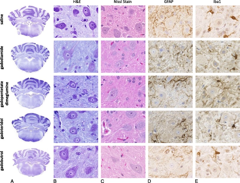
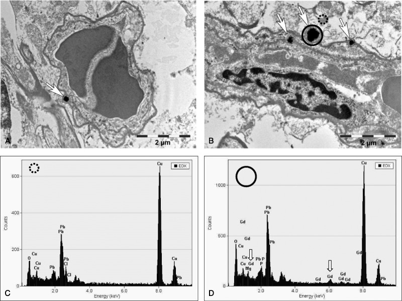
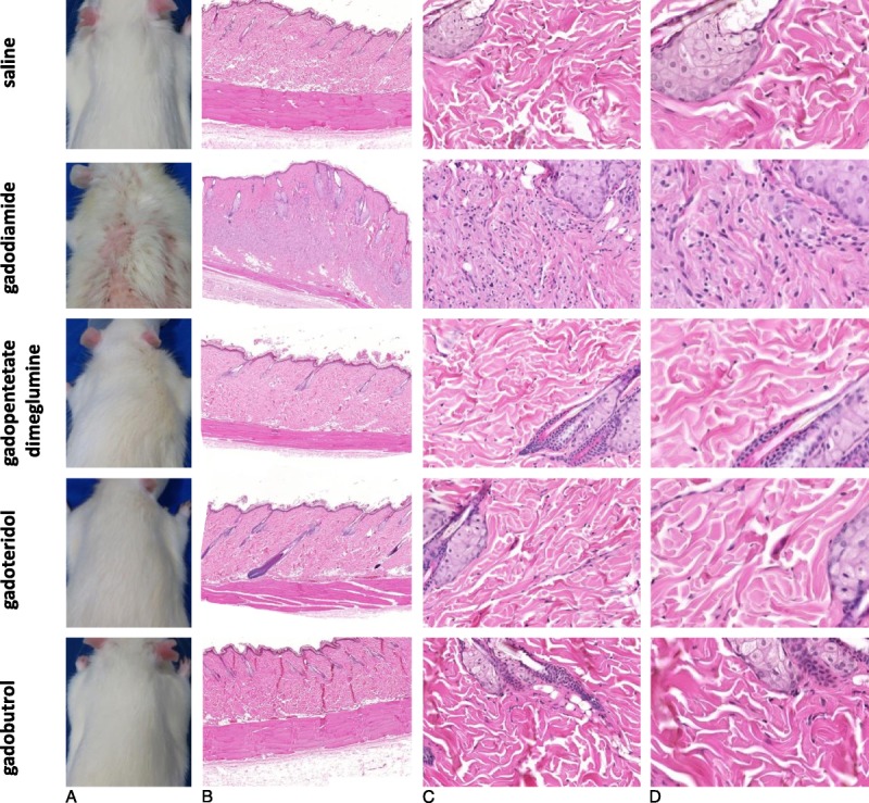
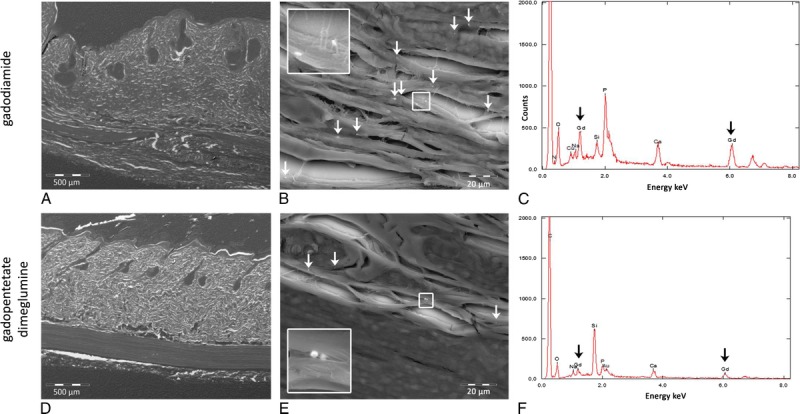
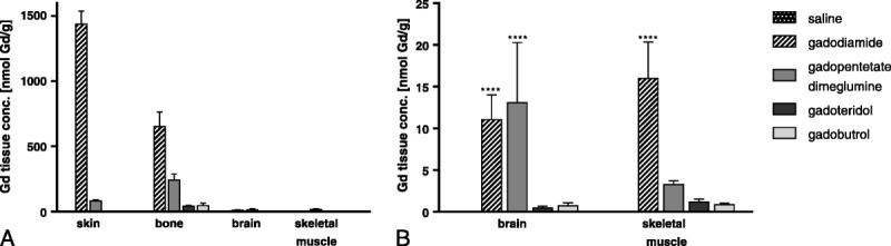
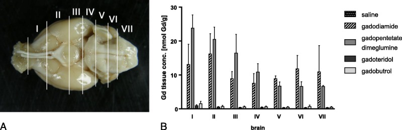
No histological changes were observed in the brain. In contrast, 4 of 10 animals in the gadodiamide group but none of the animals in other groups showed macroscopic and histological nephrogenic systemic fibrosis-like skin lesions. The Gd concentrations observed in the skin/brain samples (in nanomole Gd per gram of tissue) for each agent were as follows: gadodiamide: 1472 ± 115/11.1 ± 5.1, gadopentetate dimeglumine: 80.8 ± 6.2/13.1 ± 7.3, gadobutrol: 1.1 ± 0.5/0.7 ± 0.4, and gadoteridol: 1.7 ± 0.8/0.5 ± 0.2. The average detected residual Gd concentration in the brain was approximately 15-fold higher for linear than for macrocyclic GBCAs. The highest amounts of Gd found in brain corresponded to less than 0.0002% of the injected dose per gram of tissue. Using LA-ICP-MS, high Gd concentrations in the deep cerebellar nuclei and in the granular layer of the cerebellar cortex were detected only for linear gadodiamide and gadopentetate dimeglumine but not for gadoteridol or gadobutrol. The energy dispersive x-ray spectroscopy analysis revealed Gd-containing spots in the skin of animals administered gadodiamide and gadopentetate dimeglumine. Transmission electron microscopy revealed several Gd-containing spots in the region of the dentate nuclei in the brain of 1 animal injected with gadodiamide.
After repeated high dosing, nephrogenic systemic fibrosis-like macroscopic and histopathological lesions of the skin were observed only in some of the gadodiamide-treated animals. No histopathological findings were detected in the rodent brain. The administration of linear GBCAs was associated with significantly higher Gd concentrations in the brain and skin compared with macrocyclic GBCA administration. The results of LA-ICP-MS demonstrated local accumulation of Gd within the deep cerebellar nuclei and the granular layer only after the administration of linear agents. In summary, the detected low Gd concentrations in the skin and brain were well correlated with the higher kinetic stability of macrocyclic GBCA.
对原发性脑肿瘤或其他中枢神经系统疾病患者的回顾性研究以及尸检研究表明,在多次给予主要为线性钆基造影剂(GBCA)后,钆(Gd)会沉积在齿状核(DN)和苍白球(GP)中。然而,这种沉积并未与任何不良反应或组织病理学改变相关联。这项临床前研究的目的是系统地研究线性和大环GBCA在诱导脑和皮肤组织学变化(包括高空间分辨率下的Gd分布)方面的差异。
50只雄性Wistar-Han大鼠被随机分为对照组(生理盐水,n = 10只大鼠)和4个GBCA组(线性GBCA:钆双胺和钆喷酸葡甲胺,大环GBCA:钆布醇和钆特醇;每组n = 10只大鼠)。动物每天接受20次静脉注射,剂量为2.5 mmol Gd/kg体重。在最后一次给予GBCA后8周,处死动物,对脑和皮肤样本进行组织病理学评估(苏木精和伊红染色;甲酚紫[Nissl]染色)并进行免疫组织化学分析。使用电感耦合等离子体质谱法(ICP-MS,n = 4)分析皮肤、骨骼、脑和骨骼肌样本中的Gd浓度。使用激光烧蚀结合ICP-MS(LA-ICP-MS,n = 3)在冷冻切片中分析脑和皮肤样本中的Gd空间分布。为了实现Gd分布的超高分辨率,分别通过扫描电子显微镜耦合能量色散X射线光谱法和透射电子显微镜对注射钆双胺或生理盐水的大鼠脑切片(n = 1)进行评估。
脑中未观察到组织学变化。相比之下,钆双胺组10只动物中有4只出现了宏观和组织学上类似肾源性系统性纤维化的皮肤病变,而其他组动物均未出现。每种试剂在皮肤/脑样本中观察到的Gd浓度(每克组织中Gd的纳摩尔数)如下:钆双胺:1472±115/11.1±5.1,钆喷酸葡甲胺:80.8±6.2/13.1±7.3,钆布醇:1.1±0.5/0.7±0.4,钆特醇:1.7±0.8/0.5±0.2。线性GBCA在脑中的平均检测到的残留Gd浓度比大环GBCA高约15倍。脑中发现的最高Gd量对应于每克组织中注射剂量的不到0.0002%。使用LA-ICP-MS,仅在给予线性钆双胺和钆喷酸葡甲胺后,在小脑深部核团和小脑皮质颗粒层中检测到高Gd浓度,而钆特醇或钆布醇组未检测到。能量色散X射线光谱分析显示,给予钆双胺和钆喷酸葡甲胺的动物皮肤中有含Gd的斑点。透射电子显微镜显示,1只注射钆双胺的动物脑齿状核区域有几个含Gd的斑点。
在重复高剂量给药后,仅在部分接受钆双胺治疗的动物中观察到类似肾源性系统性纤维化的皮肤宏观和组织病理学病变。在啮齿动物脑中未检测到组织病理学发现。与给予大环GBCA相比,给予线性GBCA后脑和皮肤中的Gd浓度明显更高。LA-ICP-MS结果表明,仅在给予线性试剂后,Gd在小脑深部核团和颗粒层中局部蓄积。总之,在皮肤和脑中检测到的低Gd浓度与大环GBCA较高的动力学稳定性密切相关。