Pulley Benjamin R, Trinh Thai Q, Bentley Jared C, Politi Joel R
Department of Orthopaedic Surgery, Mount Carmel Health System, Columbus, OH, USA.
Department of Orthopaedic Surgery, Mount Carmel Health System, Columbus, OH, USA; Orthopedic ONE, Columbus, OH, USA.
Arthroplast Today. 2015 Oct 21;1(4):93-98. doi: 10.1016/j.artd.2015.07.002. eCollection 2015 Dec.
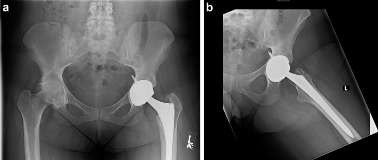
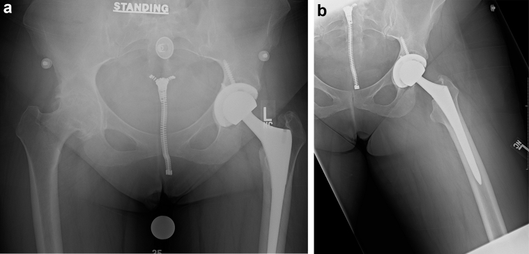
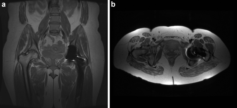
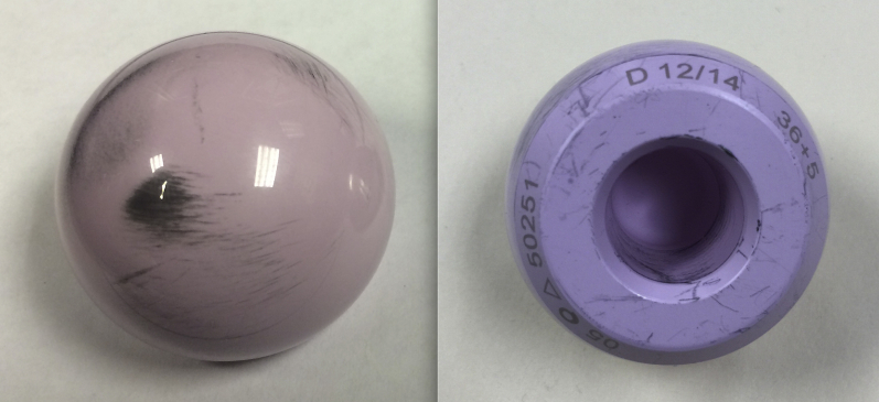
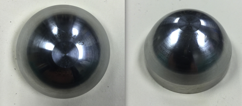
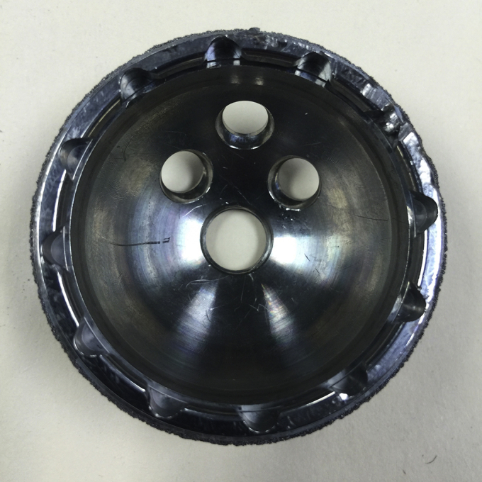
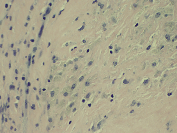
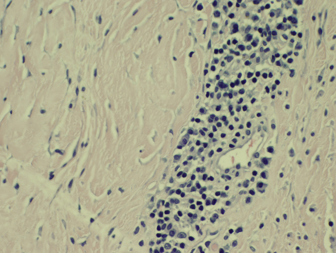
A 41-year-old woman presented 8 years after a left total hip arthroplasty. She complained of progressive groin pain for several months. Radiographs demonstrated a hard-on-hard bearing surface combination and radiolucent lines surrounding the acetabular shell. Laboratory analysis revealed a mild leukocytosis, a normal erythrocyte sedimentation rate, and a mildly elevated C-reactive protein. Serum cobalt and chromium levels were markedly elevated. Aspiration of the hip joint was negative for infection. Magnetic resonance imaging failed to demonstrate a pseudotumor. Revision total hip arthroplasty was performed, and a ceramic-on-metal bearing surface combination was explanted. Significant intraoperative findings included dark gray synovial fluid, metal transfer onto the ceramic femoral head, and a grossly loose acetabular shell pivoting about a single well-fixed screw. The explanted components otherwise appeared normal macroscopically. Histologic analysis of the capsular tissue demonstrated aseptic lymphocyte-dominated vasculitis-associated lesion and inclusion bodies consistent with third-body wear. Revision arthroplasty to a ceramic-on-polyethylene bearing surface combination was performed with a good clinical result and laboratory normalization at 9-month follow-up.
一名41岁女性在接受左侧全髋关节置换术后8年前来就诊。她主诉数月来腹股沟疼痛逐渐加重。X线片显示为硬对硬承重面组合,髋臼杯周围有透光线。实验室分析显示轻度白细胞增多、红细胞沉降率正常、C反应蛋白轻度升高。血清钴和铬水平显著升高。髋关节穿刺未发现感染。磁共振成像未显示假肿瘤。遂进行了翻修全髋关节置换术,取出了陶瓷对金属承重面组合。术中重要发现包括深灰色滑液、金属转移至陶瓷股骨头以及髋臼杯围绕一枚固定良好的螺钉严重松动。取出的假体在宏观上其他方面看起来正常。关节囊组织的组织学分析显示无菌性淋巴细胞为主的血管炎相关病变以及与三体磨损一致的包涵体。翻修为陶瓷对聚乙烯承重面组合的关节置换术,临床效果良好,9个月随访时实验室指标恢复正常。