Figueras-Roca Marc, Molins Blanca, Sala-Puigdollers Anna, Matas Jessica, Vinagre Irene, Ríos José, Adán Alfredo
Institut Clínic d'Oftalmologia (ICOF), Hospital Clínic, Barcelona, Spain.
August Pi i Sunyer Biomedical Research Institute (IDIBAPS), Barcelona, Spain.
PLoS One. 2017 Mar 22;12(3):e0173865. doi: 10.1371/journal.pone.0173865. eCollection 2017.
To study the association between peripheral blood metabolic and inflammatory factors and presence of diabetic macular edema (DME) and its related anatomic features in type 2 diabetic mellitus (T2DM) patients.
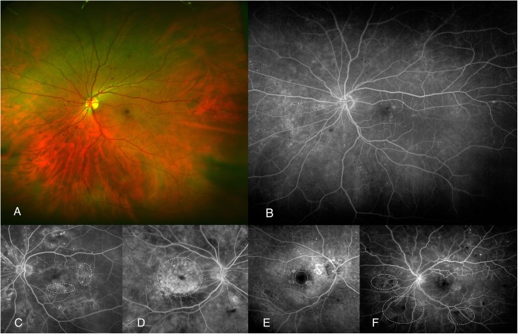
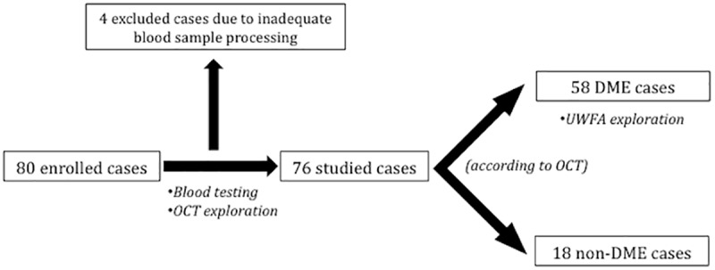
Observational cross-sectional study on a proof of concept basis. Seventy-six T2DM included patients were divided based on the presence (n = 58) or absence of DME (n = 18) according to optical coherence tomography (OCT). Ultra-widefield fluorescein angiography (UWFA) was performed in DME patients. Fasting peripheral blood sample testing included glycemia, glycated hemoglobin, creatinin and lipid levels among others. Serum levels of a broad panel of cytokines and inflammatory mediators were also analysed. OCT findings included central subfoveal thickness, diffuse retinal thickness (DRT), cystoid macular edema (CME), serous retinal detachment and epirretinal membrane. UWFA items included pattern of DME, presence of peripheral retinal ischemia and enlarged foveal avascular zone (FAZ).
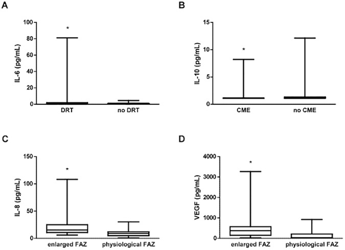
Metabolic and inflammatory factors did not statistically differ between groups. However, several inflammatory mediators did associate to certain ocular items of DME cases: IL-6 was significantly higher in patients with DRT (p = 0.044), IL-10 was decreased in patients with CME (p = 0.012), and higher IL-8 (p = 0.031) and VEGF levels (p = 0.031) were observed in patients with enlarged FAZ.
Inflammatory and metabolic peripheral blood factors in T2DM may not be differentially associated to DME when compared to non-DME cases. However, some OCT and UWFA features of DME such as DRT, CME and enlarged FAZ may be associated to certain systemic inflammatory mediators.
研究2型糖尿病(T2DM)患者外周血代谢和炎症因子与糖尿病性黄斑水肿(DME)的存在及其相关解剖学特征之间的关联。
基于概念验证的观察性横断面研究。根据光学相干断层扫描(OCT)结果,将76例纳入研究的T2DM患者分为有DME组(n = 58)和无DME组(n = 18)。对DME患者进行超广角荧光素血管造影(UWFA)检查。空腹外周血样本检测包括血糖、糖化血红蛋白、肌酐和血脂水平等。还分析了一系列细胞因子和炎症介质的血清水平。OCT检查结果包括中心凹下厚度、弥漫性视网膜厚度(DRT)、黄斑囊样水肿(CME)、浆液性视网膜脱离和视网膜前膜。UWFA检查项目包括DME的模式、周边视网膜缺血的存在以及中心凹无血管区(FAZ)扩大。
两组之间的代谢和炎症因子在统计学上无差异。然而,一些炎症介质确实与DME病例的某些眼部项目相关:DRT患者的IL-6显著更高(p = 0.044),CME患者的IL-10降低(p = 0.012),FAZ扩大的患者中观察到更高的IL-8(p = 0.031)和VEGF水平(p = 0.031)。
与无DME的病例相比,T2DM患者外周血中的炎症和代谢因子与DME的关联可能无差异。然而,DME的一些OCT和UWFA特征,如DRT、CME和FAZ扩大,可能与某些全身炎症介质相关。