Wirshing Alison C E, Cram Erin J
Department of Biology, Northeastern University, Boston, MA 02115.
Department of Biology, Northeastern University, Boston, MA 02115
Mol Biol Cell. 2017 Jul 7;28(14):1937-1949. doi: 10.1091/mbc.E17-01-0029. Epub 2017 Mar 22.
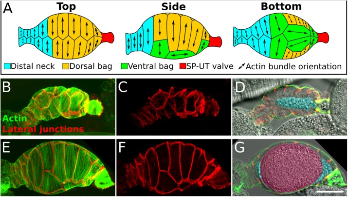
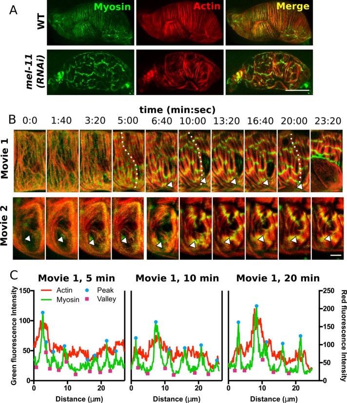
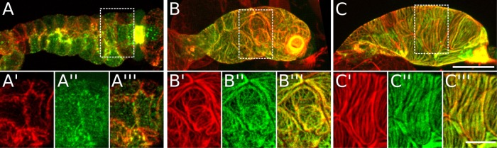
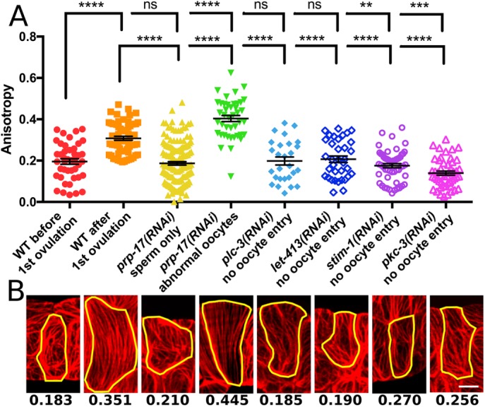
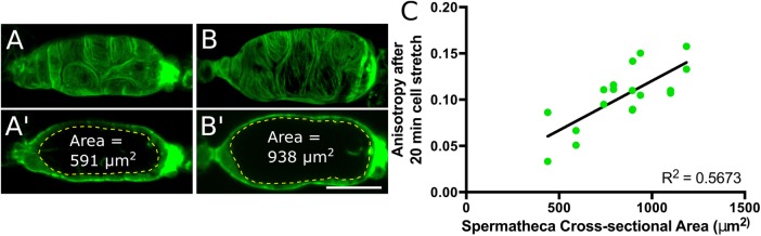
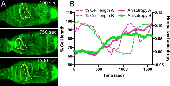
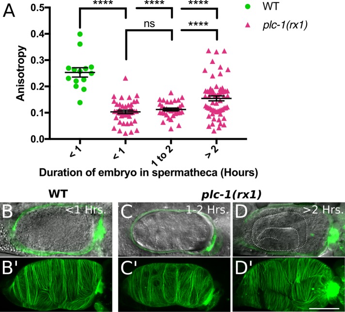
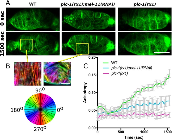
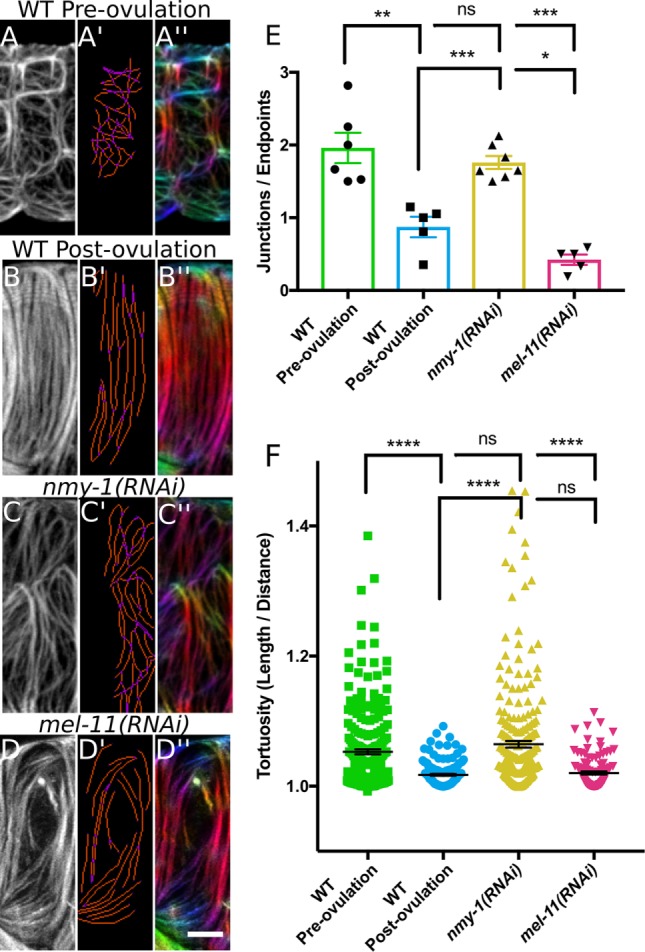
Stress fibers-contractile actomyosin bundles-are important for cellular force production and adaptation to physical stress and have been well studied within the context of cell migration. However, less is known about actomyosin bundle formation and organization in vivo and in specialized contractile cells, such as smooth muscle and myoepithelial cells. The spermatheca is a bag-like organ of 24 myoepithelial cells that houses the sperm and is the site of fertilization. During ovulation, spermathecal cells are stretched by oocyte entry and then coordinately contract to expel the fertilized embryo into the uterus. Here we use four-dimensional confocal microscopy of live animals to observe changes to spermathecal actomyosin network organization during cell stretch and contraction. Oocyte entry is required to trigger cell contraction and concomitant production of parallel actomyosin bundles. Actomyosin bundle size, connectivity, spacing, and orientation are regulated by myosin activity. We conclude that myosin drives actomyosin bundle production and that myosin activity is tightly regulated during ovulation to produce an optimally organized actomyosin network in spermathecae.
应力纤维——收缩性肌动球蛋白束——对于细胞产生力以及适应物理应力非常重要,并且在细胞迁移的背景下已经得到了充分研究。然而,关于肌动球蛋白束在体内以及在诸如平滑肌和肌上皮细胞等特殊收缩细胞中的形成和组织,我们了解得较少。受精囊是一个由24个肌上皮细胞组成的袋状器官,用于储存精子,是受精的场所。在排卵期间,受精囊细胞会因卵母细胞进入而被拉伸,然后协同收缩,将受精卵排出到子宫中。在这里,我们使用活体动物的四维共聚焦显微镜来观察受精囊肌动球蛋白网络组织在细胞拉伸和收缩过程中的变化。卵母细胞进入是触发细胞收缩以及伴随产生平行肌动球蛋白束所必需的。肌动球蛋白束的大小、连通性、间距和方向受肌球蛋白活性的调节。我们得出结论,肌球蛋白驱动肌动球蛋白束的产生,并且在排卵期间肌球蛋白活性受到严格调节,以在受精囊中产生组织优化的肌动球蛋白网络。