• 文献检索
  • 文档翻译
  • 深度研究
  • 学术资讯
  • Suppr Zotero 插件Zotero 插件
  • 邀请有礼
  • 套餐&价格
  • 历史记录
应用&插件
Suppr Zotero 插件Zotero 插件浏览器插件Mac 客户端Windows 客户端微信小程序
定价
高级版会员购买积分包购买API积分包
服务
文献检索文档翻译深度研究API 文档MCP 服务
关于我们
关于 Suppr公司介绍联系我们用户协议隐私条款
关注我们

Suppr 超能文献

核心技术专利:CN118964589B侵权必究
粤ICP备2023148730 号-1Suppr @ 2026

文献检索

告别复杂PubMed语法,用中文像聊天一样搜索,搜遍4000万医学文献。AI智能推荐,让科研检索更轻松。

立即免费搜索

文件翻译

保留排版,准确专业,支持PDF/Word/PPT等文件格式,支持 12+语言互译。

免费翻译文档

深度研究

AI帮你快速写综述,25分钟生成高质量综述,智能提取关键信息,辅助科研写作。

立即免费体验

DARC技术介绍。

An introduction to DARC technology.

作者信息

Ahmad Syed Shoeb

机构信息

Department of Ophthalmology, Queen Elizabeth Hospital, Kota Kinabalu, Malaysia.

出版信息

Saudi J Ophthalmol. 2017 Jan-Mar;31(1):38-41. doi: 10.1016/j.sjopt.2016.08.001. Epub 2016 Aug 22.

DOI:10.1016/j.sjopt.2016.08.001
PMID:28337061
原文链接:https://pmc.ncbi.nlm.nih.gov/articles/PMC5352956/
Abstract

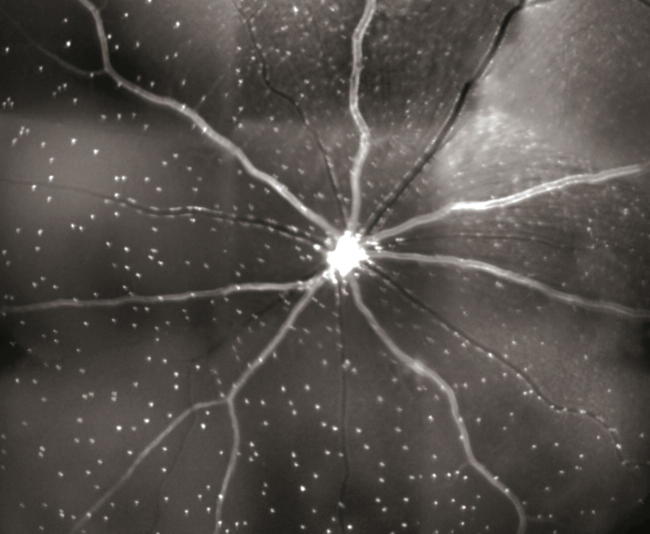
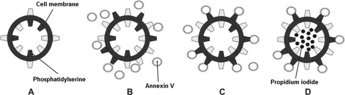
Glaucoma is a multi-factorial neurodegenerative disorder. The common denominator in all types of glaucomas is retinal ganglion cell death through apoptosis. However, this cellular demise in glaucoma is detected late by structural or functional analyses. There can be a 10-year delay prior to the appearance of visual field defects and pre-perimetric glaucoma is an issue still being addressed. However, a new cutting-edge technology called detection of apoptosing retinal cells (DARC) is being developed. This technique is capable of non-invasive, real-time visualization of apoptotic changes at the cellular level. It can detect glaucomatous cell damage at a very early stage, at the moment apoptosis starts, and thus management can be initiated even prior to development of visual field changes. In future, this technique will also be able to provide conclusive evidence of the effectiveness of treatment protocol and the need for any modifications which may be required. This article aims to provide a concise review of DARC technology.

摘要

青光眼是一种多因素神经退行性疾病。所有类型青光眼的共同特征是视网膜神经节细胞通过凋亡而死亡。然而,通过结构或功能分析在青光眼患者中检测到这种细胞死亡的时间较晚。在视野缺损出现之前可能会有10年的延迟,而视野缺损前青光眼仍是一个有待解决的问题。然而,一种名为凋亡视网膜细胞检测(DARC)的前沿新技术正在研发中。这项技术能够在细胞水平上对凋亡变化进行非侵入性实时可视化。它可以在细胞凋亡刚开始的非常早期阶段检测到青光眼性细胞损伤,从而甚至在视野变化出现之前就可以开始治疗。未来,这项技术还将能够为治疗方案的有效性以及是否需要进行任何修改提供确凿证据。本文旨在对DARC技术进行简要综述。

https://cdn.ncbi.nlm.nih.gov/pmc/blobs/4a8b/5352956/741a7773cde9/gr2.jpg
https://cdn.ncbi.nlm.nih.gov/pmc/blobs/4a8b/5352956/646d6dfb0044/gr1.jpg
https://cdn.ncbi.nlm.nih.gov/pmc/blobs/4a8b/5352956/741a7773cde9/gr2.jpg
https://cdn.ncbi.nlm.nih.gov/pmc/blobs/4a8b/5352956/646d6dfb0044/gr1.jpg
https://cdn.ncbi.nlm.nih.gov/pmc/blobs/4a8b/5352956/741a7773cde9/gr2.jpg

相似文献

1
An introduction to DARC technology.DARC技术介绍。
Saudi J Ophthalmol. 2017 Jan-Mar;31(1):38-41. doi: 10.1016/j.sjopt.2016.08.001. Epub 2016 Aug 22.
2
A semi-automated technique for labeling and counting of apoptosing retinal cells.一种用于标记和计数凋亡视网膜细胞的半自动技术。
BMC Bioinformatics. 2014 Jun 5;15:169. doi: 10.1186/1471-2105-15-169.
3
Annexins in Glaucoma. annexins 在青光眼。
Int J Mol Sci. 2018 Apr 17;19(4):1218. doi: 10.3390/ijms19041218.
4
Real-Time Imaging of Retinal Cell Apoptosis by Confocal Scanning Laser Ophthalmoscopy and Its Role in Glaucoma.共焦扫描激光眼科显微镜对视网膜细胞凋亡的实时成像及其在青光眼中的作用
Front Neurol. 2018 May 15;9:338. doi: 10.3389/fneur.2018.00338. eCollection 2018.
5
Ophthalmic Research Lecture 2018: DARC as a Potential Surrogate Marker.2018 年眼科研究讲座:DARC 作为潜在替代标志物。
Ophthalmic Res. 2020;63(1):1-7. doi: 10.1159/000500527. Epub 2019 Aug 9.
6
Real-Time Imaging of Retinal Ganglion Cell Apoptosis.视网膜神经节细胞凋亡的实时成像
Cells. 2018 Jun 15;7(6):60. doi: 10.3390/cells7060060.
7
Detecting retinal cell stress and apoptosis with DARC: Progression from lab to clinic.利用DARC检测视网膜细胞应激和凋亡:从实验室到临床的进展。
Prog Retin Eye Res. 2022 Jan;86:100976. doi: 10.1016/j.preteyeres.2021.100976. Epub 2021 Jun 5.
8
The potential of annexin-labelling for the diagnosis and follow-up of glaucoma. Annexin 标记在青光眼诊断和随访中的潜力。
Cell Tissue Res. 2013 Aug;353(2):279-85. doi: 10.1007/s00441-013-1554-5. Epub 2013 Feb 1.
9
[Aiming for zero blindness].追求零失明
Nippon Ganka Gakkai Zasshi. 2015 Mar;119(3):168-93; discussion 194.
10
Advances in retinal ganglion cell imaging.视网膜神经节细胞成像的进展。
Eye (Lond). 2015 Oct;29(10):1260-9. doi: 10.1038/eye.2015.154. Epub 2015 Aug 21.

引用本文的文献

1
From Cellular to Metabolic: Advances in Imaging of Inherited Retinal Diseases.从细胞层面到代谢层面:遗传性视网膜疾病成像技术的进展
Diagnostics (Basel). 2024 Dec 26;15(1):28. doi: 10.3390/diagnostics15010028.
2
Early diagnostics and interventional glaucoma.早期诊断与介入性青光眼
Ther Adv Ophthalmol. 2024 Oct 13;16:25158414241287431. doi: 10.1177/25158414241287431. eCollection 2024 Jan-Dec.
3
Review: Emerging Eye-Based Diagnostic Technologies for Traumatic Brain Injury.综述:创伤性脑损伤的新兴眼部诊断技术。

本文引用的文献

1
Current Concepts in the Biochemical Mechanisms of Glaucomatous Neurodegeneration.青光眼性神经变性生化机制的当前概念
J Curr Glaucoma Pract. 2013 May-Aug;7(2):49-53. doi: 10.5005/jp-journals-10008-1137. Epub 2013 May 9.
2
A semi-automated technique for labeling and counting of apoptosing retinal cells.一种用于标记和计数凋亡视网膜细胞的半自动技术。
BMC Bioinformatics. 2014 Jun 5;15:169. doi: 10.1186/1471-2105-15-169.
3
The potential of annexin-labelling for the diagnosis and follow-up of glaucoma. Annexin 标记在青光眼诊断和随访中的潜力。
IEEE Rev Biomed Eng. 2023;16:530-559. doi: 10.1109/RBME.2022.3161352. Epub 2023 Jan 5.
4
retinal imaging in translational regenerative research.转化性再生研究中的视网膜成像
Ann Transl Med. 2020 Sep;8(17):1096. doi: 10.21037/atm-20-4355.
5
Advances in Retinal Optical Imaging.视网膜光学成像的进展
Photonics. 2018 Jun;5(2). doi: 10.3390/photonics5020009. Epub 2018 Apr 27.
6
Glaucoma suspects: A practical approach.青光眼疑似病例:一种实用方法。
Taiwan J Ophthalmol. 2018 Apr-Jun;8(2):74-81. doi: 10.4103/tjo.tjo_106_17.
7
Regenerative medicine in the retina: from stem cells to cell replacement therapy.视网膜再生医学:从干细胞到细胞替代疗法。
Ther Adv Ophthalmol. 2018 Apr 26;10:2515841418774433. doi: 10.1177/2515841418774433. eCollection 2018 Jan-Dec.
Cell Tissue Res. 2013 Aug;353(2):279-85. doi: 10.1007/s00441-013-1554-5. Epub 2013 Feb 1.
4
Interference of the apoptotic signaling pathway in RGC stress response by SP600125 in moderate ocular hypertensive rats.SP600125对中度高眼压大鼠视网膜神经节细胞应激反应中凋亡信号通路的干扰作用
Chin J Physiol. 2011 Apr 30;54(2):124-32.
5
Imaging apoptosis in the eye.眼部细胞凋亡的影像学检测。
Eye (Lond). 2011 May;25(5):545-53. doi: 10.1038/eye.2011.64. Epub 2011 Mar 25.
6
Imaging multiple phases of neurodegeneration: a novel approach to assessing cell death in vivo.成像神经退行性变的多个阶段:评估体内细胞死亡的新方法。
Cell Death Dis. 2010;1(1):e3. doi: 10.1038/cddis.2009.3.
7
Mechanisms of retinal ganglion cell injury and defense in glaucoma.青光眼中原发性神经节细胞损伤和防御的机制。
Exp Eye Res. 2010 Jul;91(1):48-53. doi: 10.1016/j.exer.2010.04.002. Epub 2010 Apr 13.
8
Real-time in vivo imaging of retinal cell apoptosis after laser exposure.激光照射后视网膜细胞凋亡的实时体内成像。
Invest Ophthalmol Vis Sci. 2008 Jun;49(6):2773-80. doi: 10.1167/iovs.07-1335. Epub 2008 Feb 15.
9
Assessment of rat and mouse RGC apoptosis imaging in vivo with different scanning laser ophthalmoscopes.使用不同的扫描激光检眼镜对大鼠和小鼠视网膜神经节细胞凋亡进行体内成像评估。
Curr Eye Res. 2007 Oct;32(10):851-61. doi: 10.1080/02713680701585872.
10
Apoptosis: a review of programmed cell death.细胞凋亡:程序性细胞死亡综述
Toxicol Pathol. 2007 Jun;35(4):495-516. doi: 10.1080/01926230701320337.