Lee Jong-Geol, Shim Sehwan, Kim Min-Jung, Myung Jae Kyung, Jang Won-Suk, Bae Chang-Hwan, Lee Sun-Joo, Kim Kyeong Min, Jin Young-Woo, Lee Seung-Sook, Park Sunhoo
Laboratory of Radiation Exposure & Therapeutics, National Radiation Emergency Medical Center, KIRAMS, Seoul, Republic of Korea.
Laboratory of Radiation Exposure & Therapeutics, National Radiation Emergency Medical Center, KIRAMS, Seoul, Republic of Korea; Department of Pathology, Korea Cancer Center Hospital, KIRAMS, Seoul, Republic of Korea.
Biomed Res Int. 2017;2017:1279280. doi: 10.1155/2017/1279280. Epub 2017 Feb 27.
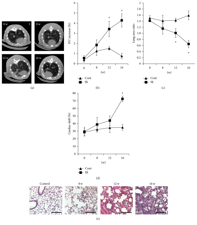
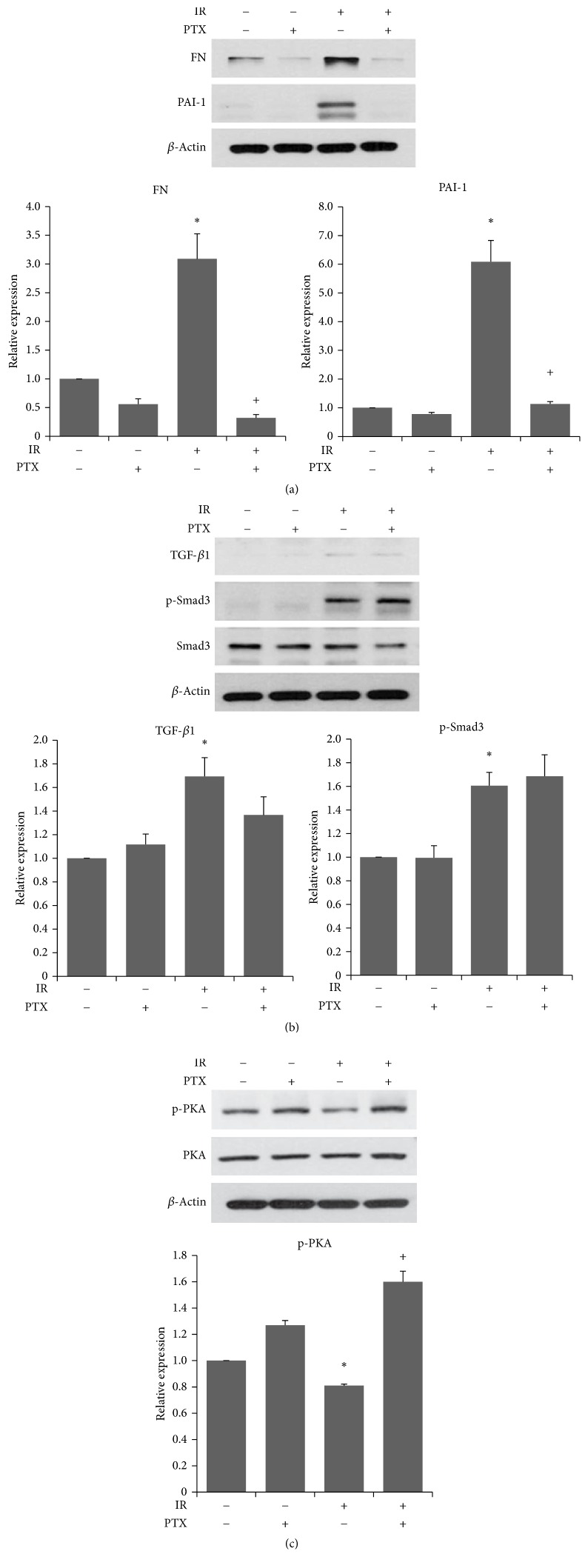
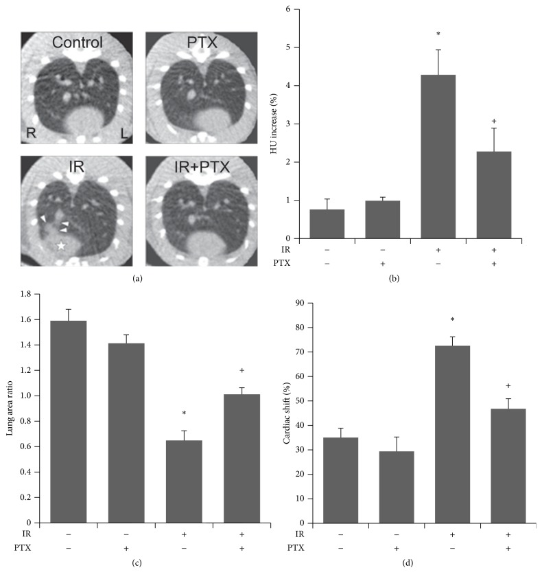
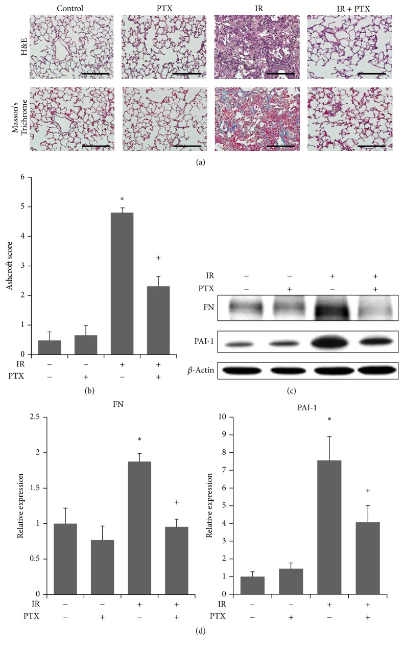
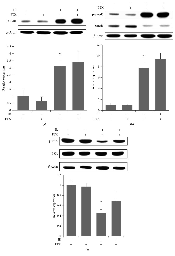
. Radiation-induced lung fibrosis (RILF) is a serious late complication of radiotherapy. In vitro studies have demonstrated that pentoxifylline (PTX) has suppressing effects in extracellular matrix production in fibroblasts, while the antifibrotic action of PTX alone using clinical dose is yet unexplored. . We used micro-computed tomography (micro-CT) and histopathological analysis to evaluate the antifibrotic effects of PTX in a rat model of RILF. . Micro-CT findings showed that lung density, volume loss, and mediastinal shift are significantly increased at 16 weeks after irradiation. Simultaneously, histological analysis demonstrated thickening of alveolar walls, destruction of alveolar structures, and excessive collagen deposition in the irradiated lung. PTX treatment effectively attenuated the fibrotic changes based on both micro-CT and histopathological analyses. Western analysis also revealed increased levels of plasminogen activator inhibitor- (PAI-) 1 and fibronectin (FN) and PTX treatment reduced expression of PAI-1 and FN by restoring protein kinase A (PKA) phosphorylation but not TGF-/Smad in both irradiated lung tissues and epithelial cells. . Our results demonstrate the antifibrotic effect of PTX on radiation-induced lung fibrosis and its effect on modulation of PKA and PAI-1 expression as possible antifibrotic mechanisms.
放射性肺纤维化(RILF)是放疗严重的晚期并发症。体外研究表明,己酮可可碱(PTX)对成纤维细胞的细胞外基质产生具有抑制作用,然而使用临床剂量的PTX单独的抗纤维化作用尚未得到探索。我们使用微型计算机断层扫描(micro-CT)和组织病理学分析来评估PTX在RILF大鼠模型中的抗纤维化作用。Micro-CT结果显示,照射后16周时肺密度、体积减少和纵隔移位显著增加。同时,组织学分析表明照射肺组织中肺泡壁增厚、肺泡结构破坏以及胶原过度沉积。基于micro-CT和组织病理学分析,PTX治疗有效减轻了纤维化改变。蛋白质印迹分析还显示,在照射的肺组织和上皮细胞中,纤溶酶原激活物抑制剂-1(PAI-1)和纤连蛋白(FN)水平升高,PTX治疗通过恢复蛋白激酶A(PKA)磷酸化而非转化生长因子/ Smad信号通路降低了PAI-1和FN的表达。我们的结果证明了PTX对放射性肺纤维化的抗纤维化作用及其对PKA和PAI-1表达调节的作用,这可能是其抗纤维化机制。