Sochacki Kem A, Dickey Andrea M, Strub Marie-Paule, Taraska Justin W
National Heart, Lung, and Blood Institute, National Institutes of Health, Bethesda, Maryland 20892, USA.
Nat Cell Biol. 2017 Apr;19(4):352-361. doi: 10.1038/ncb3498. Epub 2017 Mar 27.
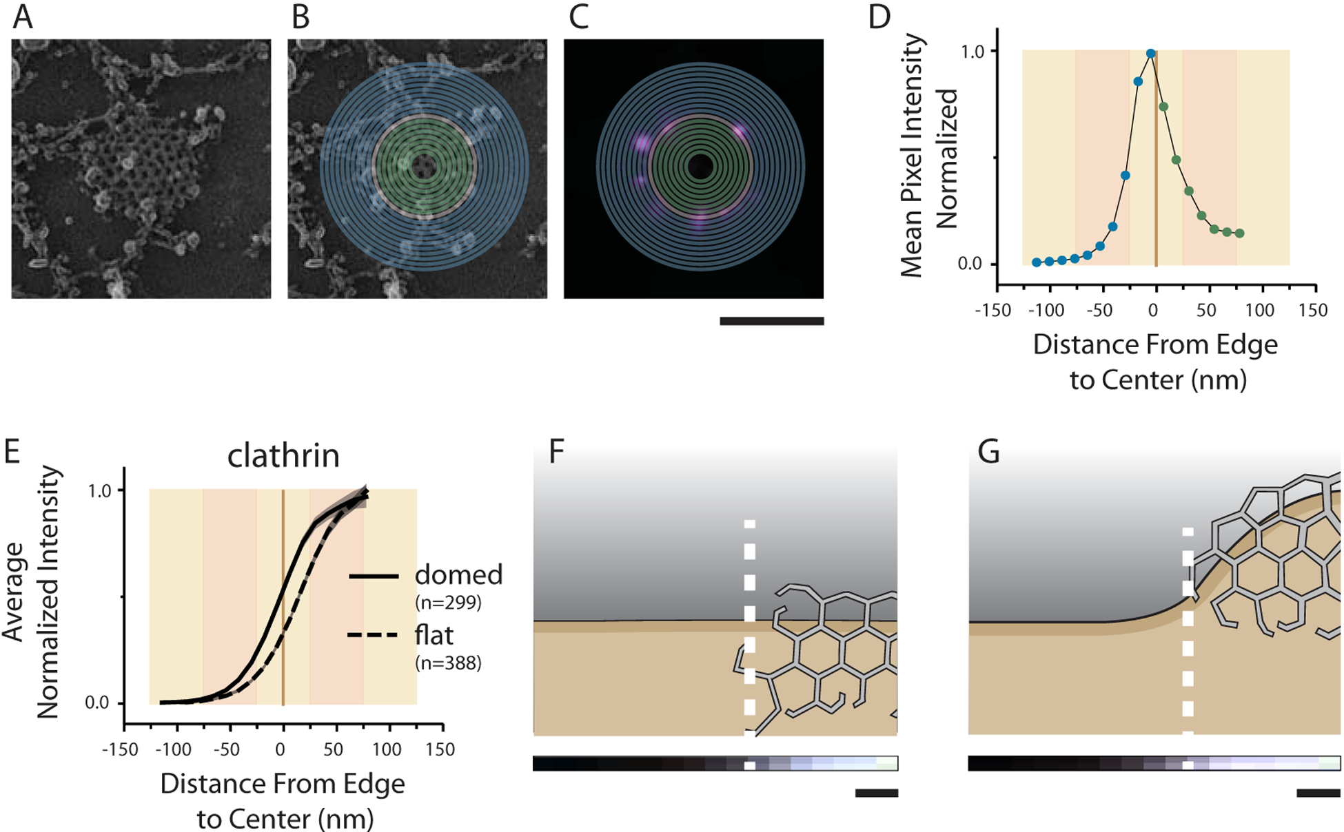
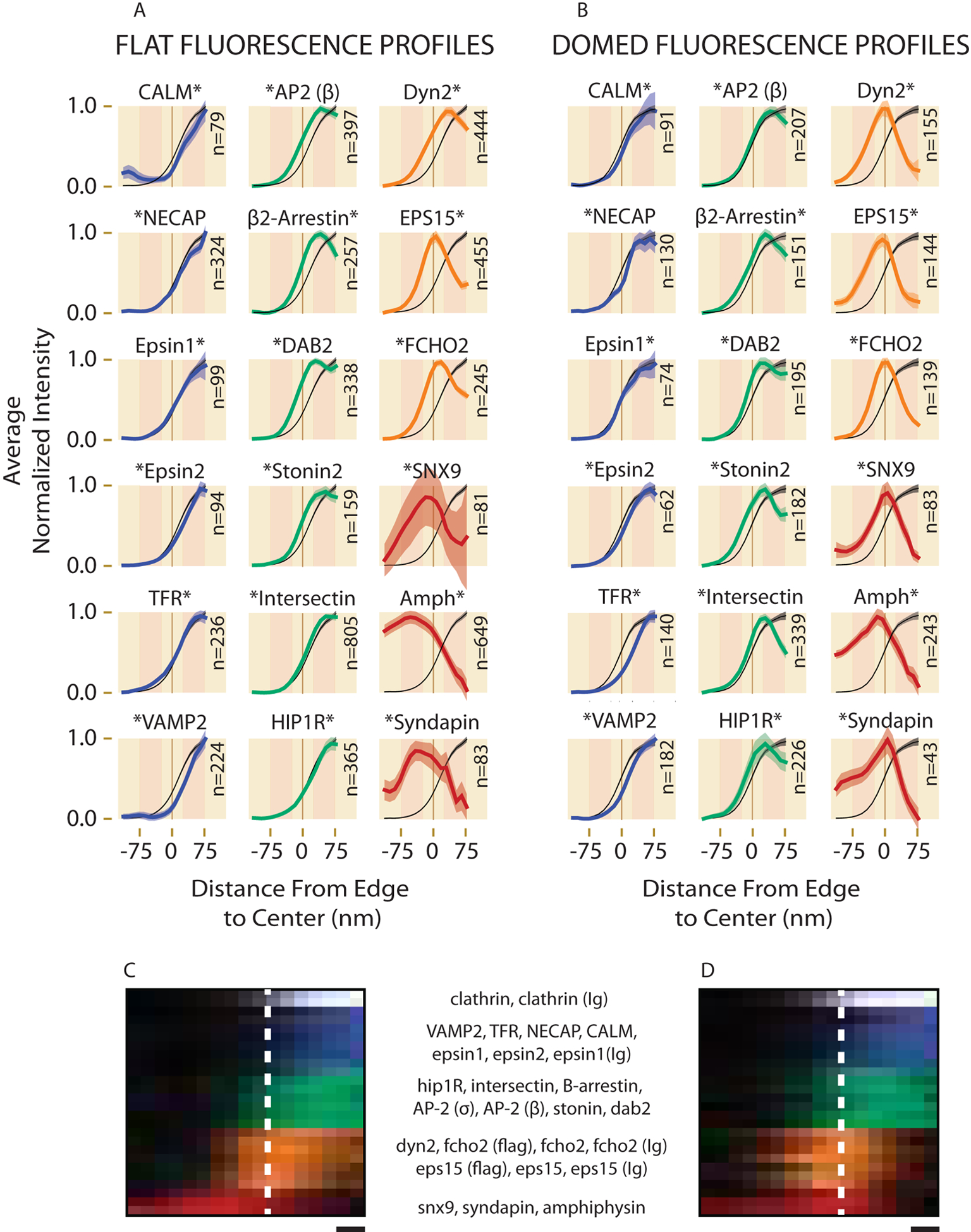
Dozens of proteins capture, polymerize and reshape the clathrin lattice during clathrin-mediated endocytosis (CME). How or if this ensemble of proteins is organized in relation to the clathrin coat is unknown. Here, we map key molecules involved in CME at the nanoscale using correlative super-resolution light and transmission electron microscopy. We localize 19 different endocytic proteins (amphiphysin1, AP2, β2-arrestin, CALM, clathrin, DAB2, dynamin2, EPS15, epsin1, epsin2, FCHO2, HIP1R, intersectin, NECAP, SNX9, stonin2, syndapin2, transferrin receptor, VAMP2) on thousands of individual clathrin structures, generating a comprehensive molecular architecture of endocytosis with nanoscale precision. We discover that endocytic proteins distribute into distinct spatial zones in relation to the edge of the clathrin lattice. The presence or concentrations of proteins within these zones vary at distinct stages of organelle development. We propose that endocytosis is driven by the recruitment, reorganization and loss of proteins within these partitioned nanoscale zones.
在网格蛋白介导的内吞作用(CME)过程中,数十种蛋白质捕获、聚合并重塑网格蛋白晶格。目前尚不清楚这组蛋白质是如何组织的,或者它们与网格蛋白包被之间是否存在关联。在这里,我们使用相关超分辨率光学显微镜和透射电子显微镜在纳米尺度上绘制了参与CME的关键分子图谱。我们在数千个单独的网格蛋白结构上定位了19种不同的内吞蛋白(发动蛋白1、衔接蛋白AP2、β2-抑制蛋白、钙网蛋白、网格蛋白、Disabled-2蛋白、发动蛋白2、表皮生长因子受体底物-15、epsin1、epsin2、FCH结构域蛋白2、亨廷顿相互作用蛋白1相关蛋白、交叉蛋白、N-乙基马来酰亚胺敏感因子附着蛋白受体相关蛋白、分选衔接蛋白9、Stonin 2、syndapin2、转铁蛋白受体、囊泡相关膜蛋白2),以纳米级精度生成了内吞作用的全面分子结构。我们发现,内吞蛋白相对于网格蛋白晶格的边缘分布在不同的空间区域。在细胞器发育的不同阶段,这些区域内蛋白质的存在或浓度各不相同。我们提出,内吞作用是由这些分隔的纳米级区域内蛋白质的募集、重组和缺失驱动的。