Boston Children's Hospital and Harvard Medical School, Department of Radiology, Boston, MA, 02115, USA.
Boston Children's Hospital and Harvard Medical School, Department of Neurology, Boston, MA, 02115, USA.
Sci Rep. 2017 Mar 28;7(1):476. doi: 10.1038/s41598-017-00525-w.
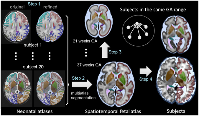
Longitudinal characterization of early brain growth in-utero has been limited by a number of challenges in fetal imaging, the rapid change in size, shape and volume of the developing brain, and the consequent lack of suitable algorithms for fetal brain image analysis. There is a need for an improved digital brain atlas of the spatiotemporal maturation of the fetal brain extending over the key developmental periods. We have developed an algorithm for construction of an unbiased four-dimensional atlas of the developing fetal brain by integrating symmetric diffeomorphic deformable registration in space with kernel regression in age. We applied this new algorithm to construct a spatiotemporal atlas from MRI of 81 normal fetuses scanned between 19 and 39 weeks of gestation and labeled the structures of the developing brain. We evaluated the use of this atlas and additional individual fetal brain MRI atlases for completely automatic multi-atlas segmentation of fetal brain MRI. The atlas is available online as a reference for anatomy and for registration and segmentation, to aid in connectivity analysis, and for groupwise and longitudinal analysis of early brain growth.
在胎儿成像中存在一些挑战,如胎儿大脑的快速生长、大小、形状和体积的变化,以及缺乏适合的胎儿大脑图像分析算法等,这限制了对胎儿大脑早期生长的纵向描述。因此,需要一个改进的胎儿大脑时空成熟的数字化脑图谱,该图谱应涵盖关键的发育阶段。我们开发了一种算法,通过在空间中进行对称的可变形配准和在年龄上进行核回归,来构建一个无偏的、四维的发育中胎儿大脑图谱。我们将该新算法应用于从 19 至 39 孕周的 81 例正常胎儿的 MRI 中构建一个时空图谱,并标记了发育中大脑的结构。我们评估了使用该图谱和其他的个体胎儿大脑 MRI 图谱,对胎儿大脑 MRI 进行全自动多图谱分割。该图谱可在线获取,可作为解剖学、配准和分割、连接分析以及早期大脑生长的群组和纵向分析的参考。