• 文献检索
  • 文档翻译
  • 深度研究
  • 学术资讯
  • Suppr Zotero 插件Zotero 插件
  • 邀请有礼
  • 套餐&价格
  • 历史记录
应用&插件
Suppr Zotero 插件Zotero 插件浏览器插件Mac 客户端Windows 客户端微信小程序
定价
高级版会员购买积分包购买API积分包
服务
文献检索文档翻译深度研究API 文档MCP 服务
关于我们
关于 Suppr公司介绍联系我们用户协议隐私条款
关注我们

Suppr 超能文献

核心技术专利:CN118964589B侵权必究
粤ICP备2023148730 号-1Suppr @ 2026

文献检索

告别复杂PubMed语法,用中文像聊天一样搜索,搜遍4000万医学文献。AI智能推荐,让科研检索更轻松。

立即免费搜索

文件翻译

保留排版,准确专业,支持PDF/Word/PPT等文件格式,支持 12+语言互译。

免费翻译文档

深度研究

AI帮你快速写综述,25分钟生成高质量综述,智能提取关键信息,辅助科研写作。

立即免费体验

胎儿脑磁共振强度、组织概率和形态的时空图谱及其在分割中的应用。

A spatiotemporal atlas of MR intensity, tissue probability and shape of the fetal brain with application to segmentation.

机构信息

Biomedical Image Computing Group, University of California San Francisco, San Francisco, CA 94143, USA.

出版信息

Neuroimage. 2010 Nov 1;53(2):460-70. doi: 10.1016/j.neuroimage.2010.06.054. Epub 2010 Jun 30.

DOI:10.1016/j.neuroimage.2010.06.054
PMID:20600970
原文链接:https://pmc.ncbi.nlm.nih.gov/articles/PMC2930902/
Abstract

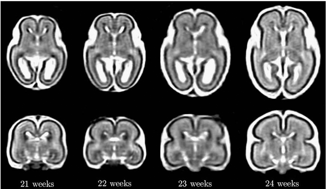
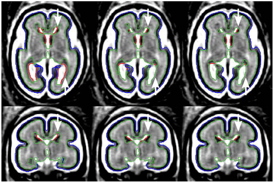
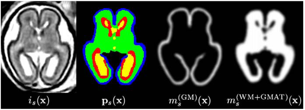
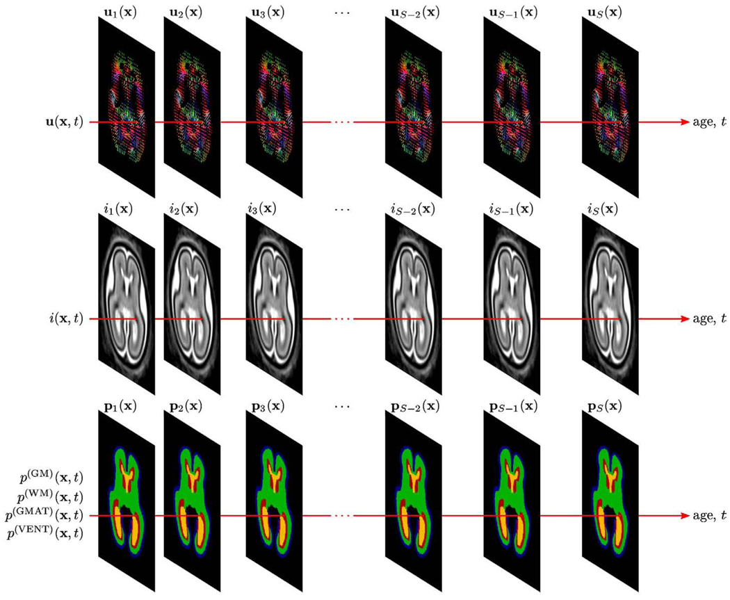
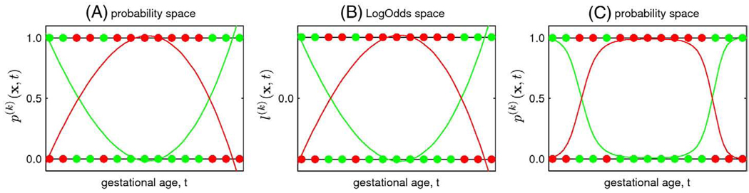
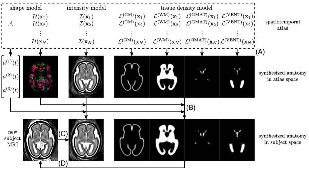
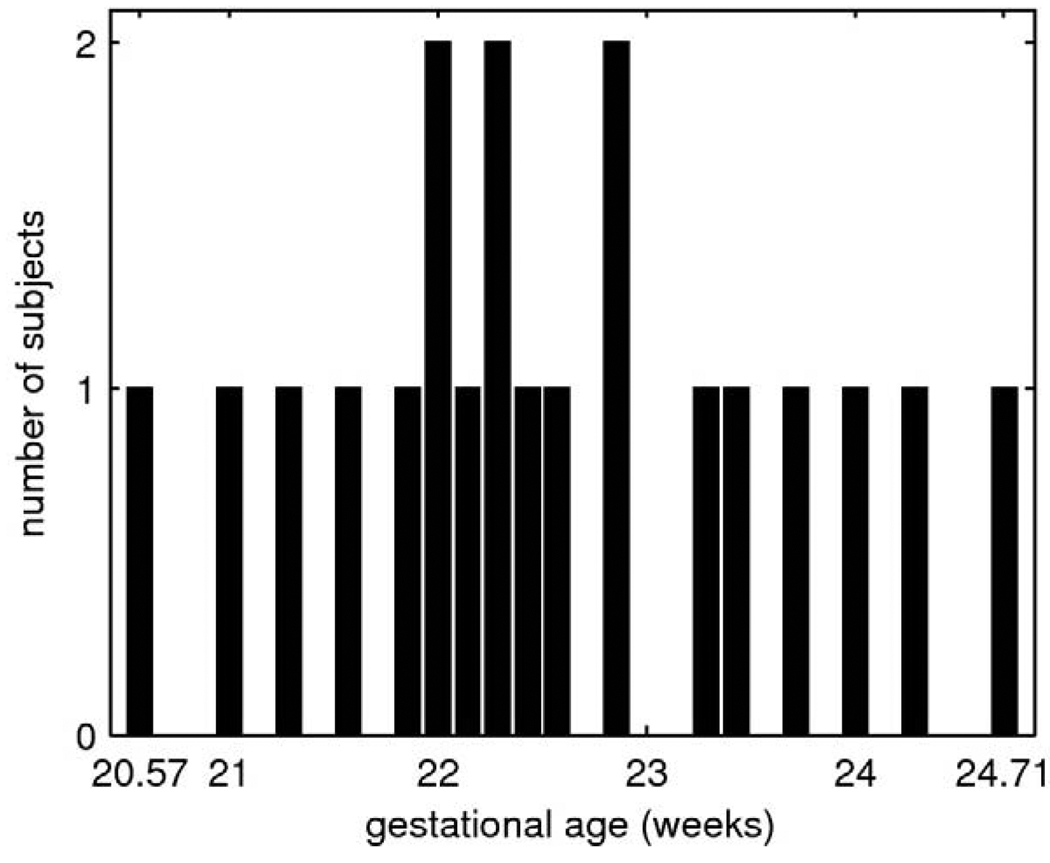
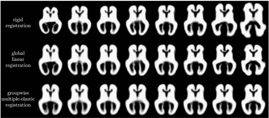
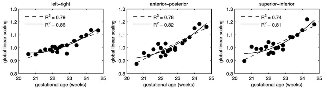
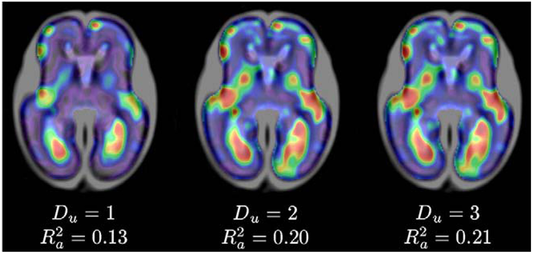
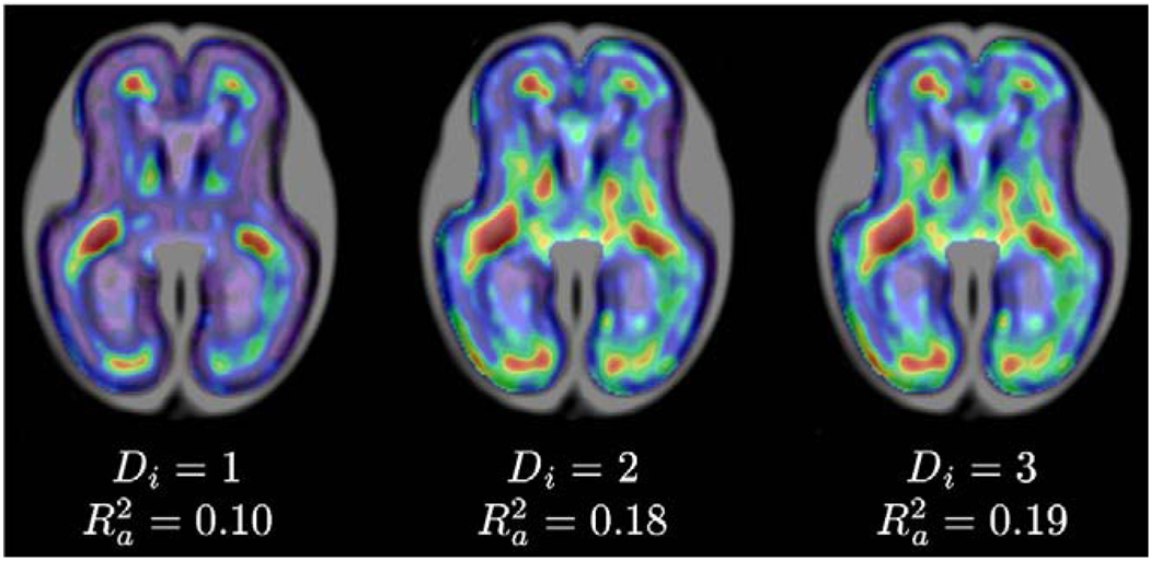
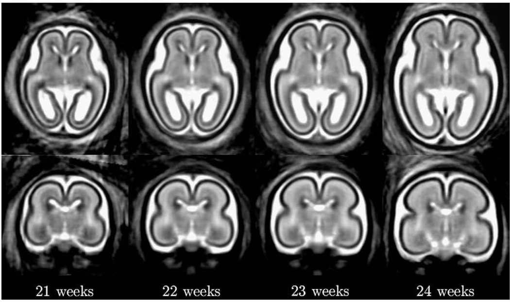
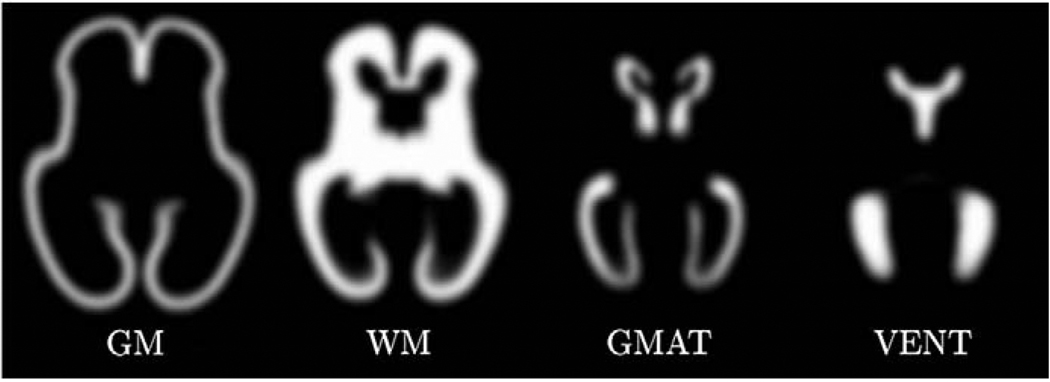
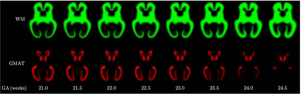
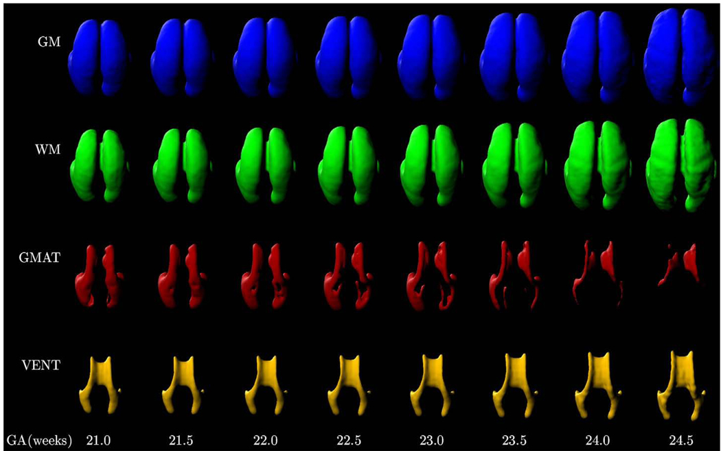
Modeling and analysis of MR images of the developing human brain is a challenge due to rapid changes in brain morphology and morphometry. We present an approach to the construction of a spatiotemporal atlas of the fetal brain with temporal models of MR intensity, tissue probability and shape changes. This spatiotemporal model is created from a set of reconstructed MR images of fetal subjects with different gestational ages. Groupwise registration of manual segmentations and voxelwise nonlinear modeling allow us to capture the appearance, disappearance and spatial variation of brain structures over time. Applying this model to atlas-based segmentation, we generate age-specific MR templates and tissue probability maps and use them to initialize automatic tissue delineation in new MR images. The choice of model parameters and the final performance are evaluated using clinical MR scans of young fetuses with gestational ages ranging from 20.57 to 24.71 weeks. Experimental results indicate that quadratic temporal models can correctly capture growth-related changes in the fetal brain anatomy and provide improvement in accuracy of atlas-based tissue segmentation.

摘要

由于人脑形态和形态测量学的快速变化,对发育中人类大脑的磁共振成像进行建模和分析是一项挑战。我们提出了一种构建胎儿大脑时空图谱的方法,该方法使用磁共振强度、组织概率和形状变化的时间模型。这个时空模型是由一组具有不同胎龄的胎儿受试者的重建磁共振图像创建的。手动分割的分组配准和体素非线性建模允许我们随时间捕获脑结构的出现、消失和空间变化。将该模型应用于基于图谱的分割,我们生成特定年龄的磁共振模板和组织概率图,并将其用于初始化新磁共振图像中的自动组织描绘。使用胎龄为 20.57 至 24.71 周的年轻胎儿的临床磁共振扫描评估模型参数的选择和最终性能。实验结果表明,二次时间模型可以正确捕获胎儿大脑解剖结构的生长相关变化,并提高基于图谱的组织分割的准确性。

https://cdn.ncbi.nlm.nih.gov/pmc/blobs/2f5c/2930902/37efbd11c680/nihms219350f15.jpg
https://cdn.ncbi.nlm.nih.gov/pmc/blobs/2f5c/2930902/65efa20be821/nihms219350f1.jpg
https://cdn.ncbi.nlm.nih.gov/pmc/blobs/2f5c/2930902/15dc086902b1/nihms219350f2.jpg
https://cdn.ncbi.nlm.nih.gov/pmc/blobs/2f5c/2930902/c6edecf5e89d/nihms219350f3.jpg
https://cdn.ncbi.nlm.nih.gov/pmc/blobs/2f5c/2930902/af4f8c5fc032/nihms219350f4.jpg
https://cdn.ncbi.nlm.nih.gov/pmc/blobs/2f5c/2930902/436209aabde2/nihms219350f5.jpg
https://cdn.ncbi.nlm.nih.gov/pmc/blobs/2f5c/2930902/2fcdf959f2d3/nihms219350f6.jpg
https://cdn.ncbi.nlm.nih.gov/pmc/blobs/2f5c/2930902/1fd388f0f251/nihms219350f7.jpg
https://cdn.ncbi.nlm.nih.gov/pmc/blobs/2f5c/2930902/9896e306aaec/nihms219350f8.jpg
https://cdn.ncbi.nlm.nih.gov/pmc/blobs/2f5c/2930902/555b6a0acb03/nihms219350f9.jpg
https://cdn.ncbi.nlm.nih.gov/pmc/blobs/2f5c/2930902/87e9e41c39eb/nihms219350f10.jpg
https://cdn.ncbi.nlm.nih.gov/pmc/blobs/2f5c/2930902/01c4ffa43616/nihms219350f11.jpg
https://cdn.ncbi.nlm.nih.gov/pmc/blobs/2f5c/2930902/1066bfa47e42/nihms219350f12.jpg
https://cdn.ncbi.nlm.nih.gov/pmc/blobs/2f5c/2930902/0631b370a556/nihms219350f13.jpg
https://cdn.ncbi.nlm.nih.gov/pmc/blobs/2f5c/2930902/82a78412f5c7/nihms219350f14.jpg
https://cdn.ncbi.nlm.nih.gov/pmc/blobs/2f5c/2930902/37efbd11c680/nihms219350f15.jpg

相似文献

1
A spatiotemporal atlas of MR intensity, tissue probability and shape of the fetal brain with application to segmentation.胎儿脑磁共振强度、组织概率和形态的时空图谱及其在分割中的应用。
Neuroimage. 2010 Nov 1;53(2):460-70. doi: 10.1016/j.neuroimage.2010.06.054. Epub 2010 Jun 30.
2
A spatio-temporal atlas of the human fetal brain with application to tissue segmentation.用于组织分割的人类胎儿脑时空图谱
Med Image Comput Comput Assist Interv. 2009;12(Pt 1):289-96. doi: 10.1007/978-3-642-04268-3_36.
3
A normative spatiotemporal MRI atlas of the fetal brain for automatic segmentation and analysis of early brain growth.胎儿大脑的规范时空 MRI 图谱,用于自动分割和分析早期脑发育。
Sci Rep. 2017 Mar 28;7(1):476. doi: 10.1038/s41598-017-00525-w.
4
Atlas-based segmentation of developing tissues in the human brain with quantitative validation in young fetuses.基于图谱的人类大脑发育组织分割及在年轻胎儿中的定量验证。
Hum Brain Mapp. 2010 Sep;31(9):1348-58. doi: 10.1002/hbm.20935.
5
Spatial-temporal atlas of human fetal brain development during the early second trimester.人类胎儿大脑在第二个妊娠中期早期的时空图谱。
Neuroimage. 2013 Nov 15;82:115-26. doi: 10.1016/j.neuroimage.2013.05.063. Epub 2013 May 31.
6
Unbiased diffeomorphic atlas construction for computational anatomy.用于计算解剖学的无偏微分同胚图谱构建
Neuroimage. 2004;23 Suppl 1:S151-60. doi: 10.1016/j.neuroimage.2004.07.068.
7
Atlas-based segmentation of the germinal matrix from in utero clinical MRI of the fetal brain.基于图谱的胎儿脑宫内临床磁共振成像中生发基质的分割
Med Image Comput Comput Assist Interv. 2008;11(Pt 1):351-8. doi: 10.1007/978-3-540-85988-8_42.
8
Automatic quantification of normal cortical folding patterns from fetal brain MRI.从胎儿脑磁共振成像中自动定量分析正常皮质折叠模式。
Neuroimage. 2014 May 1;91:21-32. doi: 10.1016/j.neuroimage.2014.01.034. Epub 2014 Jan 25.
9
Unsupervised segmentation, clustering, and groupwise registration of heterogeneous populations of brain MR images.无监督的脑磁共振图像异质人群分割、聚类和组间配准。
IEEE Trans Med Imaging. 2014 Feb;33(2):201-24. doi: 10.1109/TMI.2013.2270114. Epub 2013 Jun 19.
10
Multi-atlas segmentation of the whole hippocampus and subfields using multiple automatically generated templates.使用多个自动生成的模板对整个海马体及其子区进行多图谱分割。
Neuroimage. 2014 Nov 1;101:494-512. doi: 10.1016/j.neuroimage.2014.04.054. Epub 2014 Apr 29.

引用本文的文献

1
An MRI Atlas of the Human Fetal Brain: Reference and Segmentation Tools for Fetal Brain MRI Analysis.《人类胎儿脑部磁共振成像图谱:胎儿脑部磁共振成像分析的参考与分割工具》
ArXiv. 2025 Aug 28:arXiv:2508.15034v2.
2
Longitudinal Assessment of Abnormal Cortical Folding in Fetuses and Neonates With Isolated Non-Severe Ventriculomegaly.孤立性非重度脑室扩大胎儿及新生儿皮质折叠异常的纵向评估
Brain Behav. 2025 Jan;15(1):e70255. doi: 10.1002/brb3.70255.
3
Pathogenesis of Germinal Matrix Hemorrhage: Insights from Single-Cell Transcriptomics.生发基质出血的发病机制:来自单细胞转录组学的见解

本文引用的文献

1
Atlas-based segmentation of developing tissues in the human brain with quantitative validation in young fetuses.基于图谱的人类大脑发育组织分割及在年轻胎儿中的定量验证。
Hum Brain Mapp. 2010 Sep;31(9):1348-58. doi: 10.1002/hbm.20935.
2
MR imaging of the fetal brain.胎儿脑的磁共振成像。
Pediatr Radiol. 2010 Jan;40(1):68-81. doi: 10.1007/s00247-009-1459-3. Epub 2009 Nov 24.
3
Intersection based motion correction of multislice MRI for 3-D in utero fetal brain image formation.基于交叠的多层 MRI 运动校正用于三维宫内胎儿脑图像形成。
Annu Rev Pathol. 2025 Jan;20(1):221-243. doi: 10.1146/annurev-pathmechdis-111523-023446. Epub 2025 Jan 2.
4
Fetal brain MRI atlases and datasets: A review.胎儿脑 MRI 图谱和数据集:综述。
Neuroimage. 2024 Apr 15;292:120603. doi: 10.1016/j.neuroimage.2024.120603. Epub 2024 Apr 6.
5
Normative spatiotemporal fetal brain maturation with satisfactory development at 2 years.正常胎儿脑成熟的时空模式,2 岁时发育良好。
Nature. 2023 Nov;623(7985):106-114. doi: 10.1038/s41586-023-06630-3. Epub 2023 Oct 25.
6
A spatio-temporal atlas of the developing fetal brain with spina bifida aperta.开放性脊柱裂胎儿发育中脑的时空图谱。
Open Res Eur. 2022 Aug 31;1:123. doi: 10.12688/openreseurope.13914.2. eCollection 2021.
7
Neuromorphological Atlas of Human Prenatal Brain Development: White Paper.《人类产前脑发育神经形态学图谱:白皮书》
Life (Basel). 2023 May 13;13(5):1182. doi: 10.3390/life13051182.
8
Development of cortical folds in the human brain: An attempt to review biological hypotheses, early neuroimaging investigations and functional correlates.人类大脑脑回发育:对生物学假说、早期神经影像学研究及功能相关性的综述尝试。
Dev Cogn Neurosci. 2023 Jun;61:101249. doi: 10.1016/j.dcn.2023.101249. Epub 2023 Apr 25.
9
The nnU-Net based method for automatic segmenting fetal brain tissues.基于nnU-Net的胎儿脑组织自动分割方法。
Health Inf Sci Syst. 2023 Mar 27;11(1):17. doi: 10.1007/s13755-023-00220-3. eCollection 2023 Dec.
10
Fetal temporal sulcus depth asymmetry has prognostic value for language development.胎儿颞横回深度不对称对语言发育具有预后价值。
Commun Biol. 2023 Jan 27;6(1):109. doi: 10.1038/s42003-023-04503-z.
IEEE Trans Med Imaging. 2010 Jan;29(1):146-58. doi: 10.1109/TMI.2009.2030679. Epub 2009 Sep 9.
4
Automatic segmentation of newborn brain MRI.新生儿脑部磁共振成像的自动分割
Neuroimage. 2009 Aug 15;47(2):564-72. doi: 10.1016/j.neuroimage.2009.04.068. Epub 2009 May 3.
5
The effect of template choice on morphometric analysis of pediatric brain data.模板选择对儿科脑数据形态计量分析的影响。
Neuroimage. 2009 Apr 15;45(3):769-77. doi: 10.1016/j.neuroimage.2008.12.046. Epub 2009 Jan 6.
6
MR imaging methods for assessing fetal brain development.评估胎儿脑发育的磁共振成像方法。
Dev Neurobiol. 2008 May;68(6):700-11. doi: 10.1002/dneu.20614.
7
Tracking myocardial deformation using phase contrast MR velocity fields: a stochastic approach.使用相位对比磁共振速度场追踪心肌变形:一种随机方法。
IEEE Trans Med Imaging. 1996;15(4):453-65. doi: 10.1109/42.511749.
8
Segmentation of brain MRI in young children.幼儿脑部磁共振成像的分割
Acad Radiol. 2007 Nov;14(11):1350-66. doi: 10.1016/j.acra.2007.07.020.
9
Automatic segmentation and reconstruction of the cortex from neonatal MRI.从新生儿磁共振成像中自动分割和重建皮质。
Neuroimage. 2007 Nov 15;38(3):461-77. doi: 10.1016/j.neuroimage.2007.07.030. Epub 2007 Aug 7.
10
Using the logarithm of odds to define a vector space on probabilistic atlases.使用对数优势在概率图谱上定义向量空间。
Med Image Anal. 2007 Oct;11(5):465-77. doi: 10.1016/j.media.2007.06.003. Epub 2007 Jun 22.