Wiśniewski Marcin, Baumgart Mariusz, Grzonkowska Magdalena, Małkowski Bogdan, Wilińska-Jankowska Arnika, Siedlecki Zygmunt, Szpinda Michał
Department of Normal Anatomy, The Ludwik Rydygier Collegium Medicum in Bydgoszcz, The Nicolaus Copernicus University in Toruń, Łukasiewicza 1 Street, Bydgoszcz, 85-821, Poland.
Department of Positron Emission Tomography and Molecular Imaging, The Ludwik Rydygier Collegium Medicum in Bydgoszcz, The Nicolaus Copernicus University in Toruń, Bydgoszcz, Poland.
Surg Radiol Anat. 2017 Oct;39(10):1107-1116. doi: 10.1007/s00276-017-1849-4. Epub 2017 Mar 29.
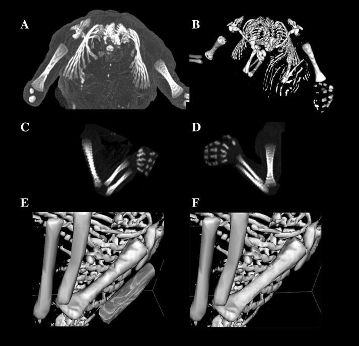
The knowledge of the development of the humeral shaft ossification center may be useful both in determining the fetal stage and maturity and for detecting congenital disorders, as well. This study was performed to quantitatively examine the humeral shaft ossification center with respect to its linear, planar, and volumetric parameters.
Using methods of CT, digital image analysis, and statistics, the size of the humeral shaft ossification center in 48 spontaneously aborted human fetuses aged 17-30 weeks was studied.
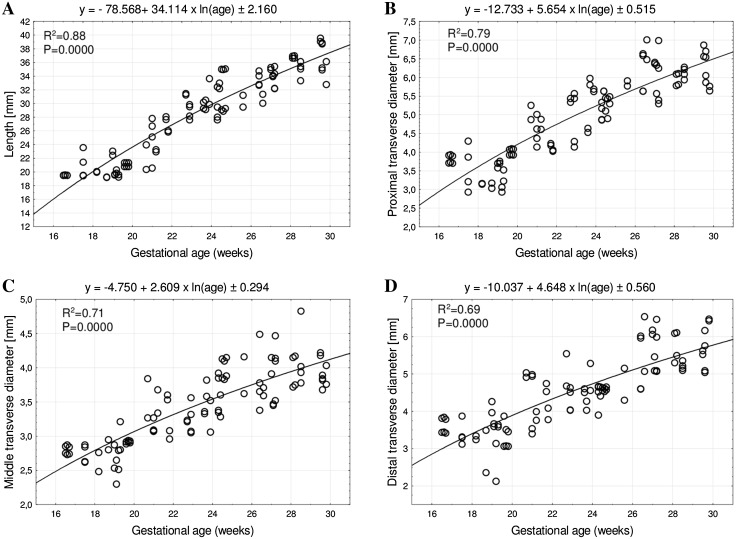
With no sex differences, the best-fit growth dynamics for the humeral shaft ossification center was modeled by the following functions: y = -78.568 + 34.114 × ln (age) ± 2.160 for its length, y = -12.733 + 5.654 × ln(age) ± 0.515 for its proximal transverse diameter, y = -4.750 + 2.609 × ln (age) ± 0.294 for its middle transverse diameter, y = -10.037 + 4.648 × ln (age) ± 0.560 for its distal transverse diameter, y = -146.601 + 11.237 × age ± 19.907 for its projection surface area, and y = 121.159 + 0.001 × (age) ± 102.944 for its volume.
With no sex differences, the ossification center of the humeral shaft grows logarithmically with respect to its length and transverse diameters, linearly with respect to its projection surface area, and fourth-degree polynomially with respect to its volume. The obtained morphometric data of the humeral shaft ossification center are considered normative for respective prenatal weeks and may be of relevance in both the estimation of fetal ages and the ultrasonic diagnostics of congenital defects.
肱骨干骨化中心发育的相关知识对于确定胎儿阶段和成熟度以及检测先天性疾病可能都很有用。本研究旨在对肱骨干骨化中心的线性、平面和体积参数进行定量研究。
采用CT、数字图像分析和统计学方法,对48例17 - 30周自然流产的人类胎儿的肱骨干骨化中心大小进行了研究。
肱骨干骨化中心的最佳拟合生长动力学模型在性别上无差异,其长度的模型为:y = -78.568 + 34.114 × ln(年龄) ± 2.160;近端横径的模型为:y = -12.733 + 5.654 × ln(年龄) ± 0.515;中间横径的模型为:y = -4.750 + 2.609 × ln(年龄) ± 0.294;远端横径的模型为:y = -10.037 + 4.648 × ln(年龄) ± 0.560;投影表面积的模型为:y = -146.601 + 11.237 × 年龄 ± 19.907;体积的模型为:y = 121.159 + 0.001 × (年龄) ± 102.944。
肱骨干骨化中心在性别上无差异,其长度和横径呈对数生长,投影表面积呈线性生长,体积呈四次多项式生长。所获得的肱骨干骨化中心形态测量数据被认为是各产前周的标准数据,可能在估计胎儿年龄和先天性缺陷的超声诊断中都具有相关性。