Li Zuowei, Zheng Xiaonan, Li Ping, Itoua Eudes Saturnin Régis, Moukassa Donatien, Ndinga Andely Françoise
Department of Integrated Traditional Chinese and Western Medicine Encephalopathy, Tianjin Nankai Hospital, Tianjin, China (mainland).
Department of Acupuncture, Tianjin Institute of Chinese Medicine, Tianjin, China (mainland).
Med Sci Monit. 2017 Mar 30;23:1522-1532. doi: 10.12659/msm.897689.
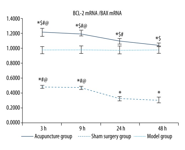
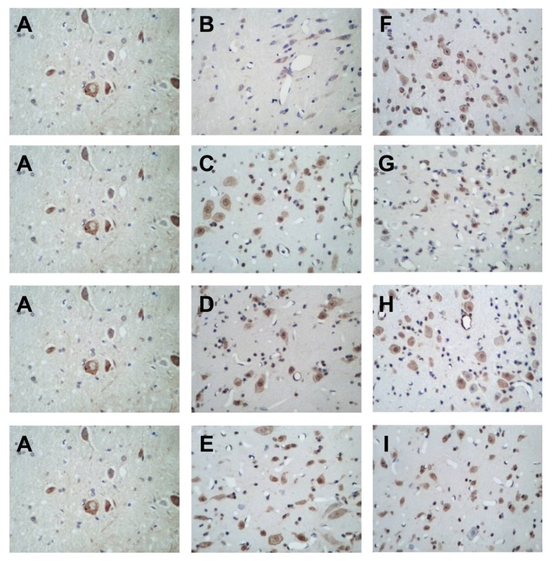
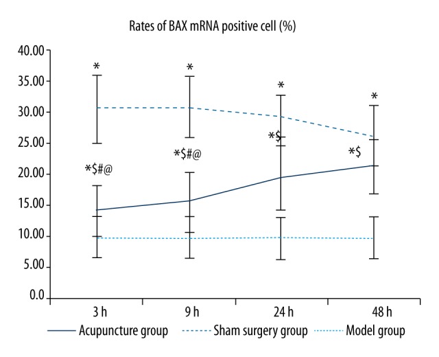
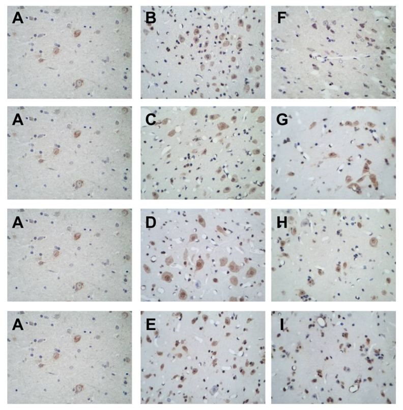
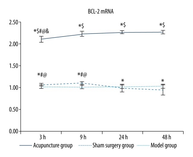
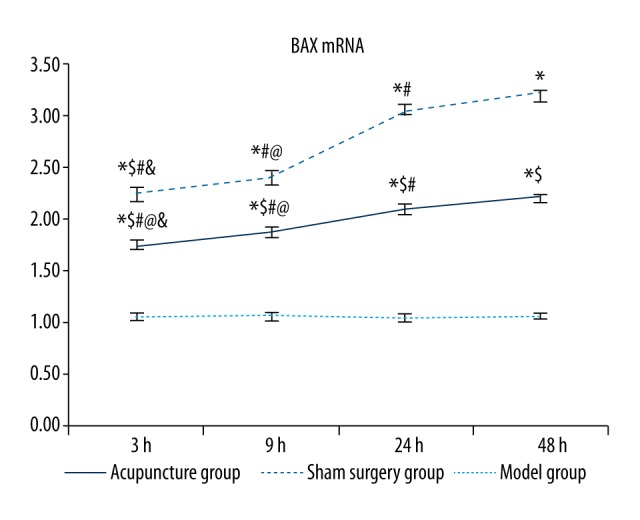
BACKGROUND To explore the time-dependent effects of acupuncture on mRNA levels of the apoptotic factors BCL-2 and BAX in a rat cerebral hemorrhage model, slow injection of autologous blood to the caudate nucleus was used to generate the cerebral hemorrhage model. MATERIAL AND METHODS A sham surgery control group, groups with acupuncture applied 3, 9, 24, and 48 hours after model induction, and time-matched model-only control groups were used. In situ hybridization was used to detect BCL-2 and BAX mRNA expression, and semi-quantitative RT-PCR was used to measure the expression. RESULTS The number of BCL-2 and BAX mRNA-positive cells significantly increased during the acute phase of cerebral hemorrhage. BCL-2 mRNA was significantly upregulated in acupuncture groups compared to other groups, whereas BAX mRNA levels in the acupuncture groups were lower in the other groups, except for the sham surgery group. Additionally, earlier acupuncture intervention was associated with a lower ratio of expression between the two genes. Changes in BCL-2 and BAX mRNA expression were consistent with changes in the number of cells positive for BCL-2 and BAX mRNA; however, the change in the expression ratio was consistent with the change in the number of cells positive for BCL-2 mRNA, but opposite to the change in the number of cells positive for BAX mRNA. CONCLUSIONS Acupuncture ameliorated changes in expression of apoptotic factors in the brain induced by acute cerebral hemorrhage and may thus protect the brain, with greater efficacy when the delay before acupuncture was minimized.
为探讨针刺对大鼠脑出血模型中凋亡因子BCL-2和BAX mRNA水平的时间依赖性影响,采用向尾状核缓慢注射自体血的方法建立脑出血模型。材料与方法:设立假手术对照组、模型诱导后3、9、24和48小时针刺组以及时间匹配的单纯模型对照组。采用原位杂交检测BCL-2和BAX mRNA表达,采用半定量RT-PCR检测其表达量。结果:脑出血急性期BCL-2和BAX mRNA阳性细胞数量显著增加。与其他组相比,针刺组BCL-2 mRNA显著上调,而针刺组BAX mRNA水平除假手术组外低于其他组。此外,早期针刺干预与两个基因的表达比值较低有关。BCL-2和BAX mRNA表达变化与BCL-2和BAX mRNA阳性细胞数量变化一致;然而,表达比值的变化与BCL-2 mRNA阳性细胞数量变化一致,但与BAX mRNA阳性细胞数量变化相反。结论:针刺可改善急性脑出血诱导的脑内凋亡因子表达变化,从而可能保护大脑,针刺延迟时间越短疗效越好。