• 文献检索
  • 文档翻译
  • 深度研究
  • 学术资讯
  • Suppr Zotero 插件Zotero 插件
  • 邀请有礼
  • 套餐&价格
  • 历史记录
应用&插件
Suppr Zotero 插件Zotero 插件浏览器插件Mac 客户端Windows 客户端微信小程序
定价
高级版会员购买积分包购买API积分包
服务
文献检索文档翻译深度研究API 文档MCP 服务
关于我们
关于 Suppr公司介绍联系我们用户协议隐私条款
关注我们

Suppr 超能文献

核心技术专利:CN118964589B侵权必究
粤ICP备2023148730 号-1Suppr @ 2026

文献检索

告别复杂PubMed语法,用中文像聊天一样搜索,搜遍4000万医学文献。AI智能推荐,让科研检索更轻松。

立即免费搜索

文件翻译

保留排版,准确专业,支持PDF/Word/PPT等文件格式,支持 12+语言互译。

免费翻译文档

深度研究

AI帮你快速写综述,25分钟生成高质量综述,智能提取关键信息,辅助科研写作。

立即免费体验

乳腺良恶性病变的鉴别:3.0T磁共振利用读出分段回波平面成像技术对乳腺病变进行扩散加权成像定性分析的附加价值

Differentiation of malignant and benign breast lesions: Added value of the qualitative analysis of breast lesions on diffusion-weighted imaging (DWI) using readout-segmented echo-planar imaging at 3.0 T.

作者信息

An Yeong Yi, Kim Sung Hun, Kang Bong Joo

机构信息

Department of Radiology, St. Vincent's Hospital, College of Medicine, The Catholic University of Korea, 93, Jungbu-daero, Paldal-gu, Suwon-si, Gyeonggi-do, Republic of Korea.

Department of Radiology, Seoul St. Mary's Hospital, College of Medicine, The Catholic University of Korea, 222, Banpo-daero, Seocho-gu, Seoul, Republic of Korea.

出版信息

PLoS One. 2017 Mar 30;12(3):e0174681. doi: 10.1371/journal.pone.0174681. eCollection 2017.

DOI:10.1371/journal.pone.0174681
PMID:28358833
原文链接:https://pmc.ncbi.nlm.nih.gov/articles/PMC5373600/
Abstract

OBJECTIVE

To determine the added value of qualitative analysis as an adjunct to quantitative analysis for the discrimination of benign and malignant lesions in patients with breast cancer using diffusion-weighted imaging (DWI) with readout-segmented echo-planar imaging (rs-EPI).

METHODS

A total of 99 patients with 144 lesions were reviewed from our prospectively collected database. DWI data were obtained using rs-EPI acquired at 3.0 T. The diagnostic performances of DWI in the qualitative, quantitative, and combination analyses were compared with that of dynamic contrast-enhanced magnetic resonance imaging (DCE-MRI). Additionally, the effect of lesion size on the diagnostic performance of the DWI combination analysis was evaluated.

RESULTS

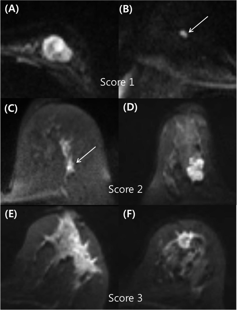
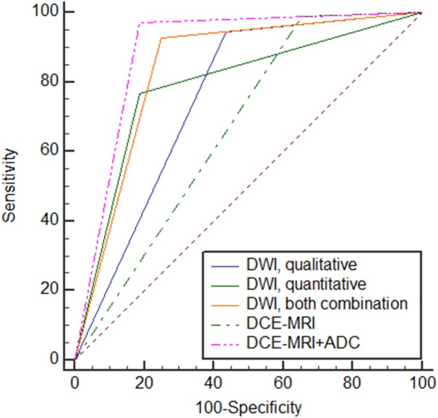
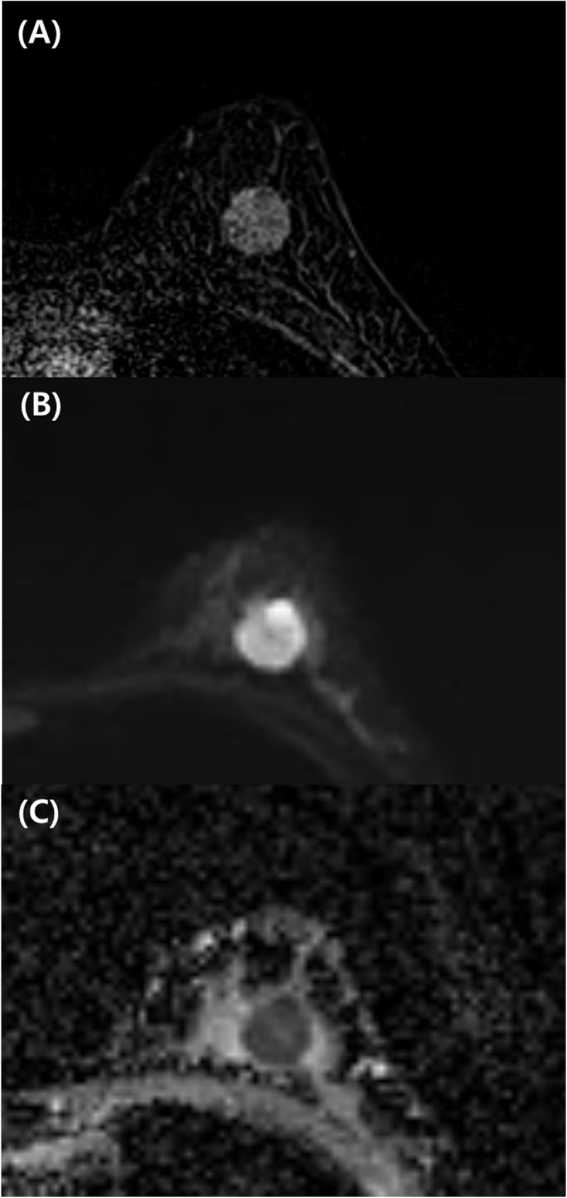
The strongest indicators of malignancy on DWI were a heterogeneous pattern (P = 0.005) and an apparent diffusion coefficient (ADC) value <1.0 × 10⁻³ mm2/sec (P = 0.002). The area under the curve (AUC) values for the qualitative analysis, quantitative analysis, and combination analysis on DWI were 0.732 (95% CI, 0.651-0.803), 0.780 (95% CI, 0.703-0.846), and 0.826 (95% CI, 0.754-0.885), respectively (P<0.0001). The AUC for the combination analysis on DWI was superior to that for DCE-MRI alone (0.651, P = 0.003) but inferior to that for DCE-MRI plus the ADC value (0.883, P = 0.03). For the DWI combination analysis, the sensitivity was significantly lower in the size ≤1 cm group than in the size >1 cm group (80% vs. 95.6%, P = 0.034).

CONCLUSIONS

Qualitative analysis of tumor morphology was diagnostically applicable on DWI using rs-EPI. This qualitative analysis adds value to quantitative analyses for lesion characterization in patients with breast cancer.

摘要

目的

利用读出分段回波平面成像(rs-EPI)的扩散加权成像(DWI),确定定性分析作为定量分析辅助手段在鉴别乳腺癌患者良性和恶性病变中的附加价值。

方法

从我们前瞻性收集的数据库中回顾了99例有144个病变的患者。使用在3.0 T下采集的rs-EPI获得DWI数据。将DWI在定性、定量和联合分析中的诊断性能与动态对比增强磁共振成像(DCE-MRI)进行比较。此外,评估了病变大小对DWI联合分析诊断性能的影响。

结果

DWI上恶性肿瘤的最强指标是不均匀模式(P = 0.005)和表观扩散系数(ADC)值<1.0×10⁻³ mm2/秒(P = 0.002)。DWI定性分析、定量分析和联合分析的曲线下面积(AUC)值分别为0.732(95% CI,0.651-0.803)、0.780(95% CI,0.703-0.846)和0.826(95% CI,0.754-0.885)(P<0.0001)。DWI联合分析的AUC优于单独的DCE-MRI(0.651,P = 0.003),但低于DCE-MRI加ADC值(0.883,P = 0.03)。对于DWI联合分析,大小≤1 cm组的敏感性显著低于大小>1 cm组(80%对95.6%,P = 0.034)。

结论

使用rs-EPI对DWI进行肿瘤形态的定性分析在诊断上是适用的。这种定性分析为乳腺癌患者病变特征的定量分析增加了价值。

https://cdn.ncbi.nlm.nih.gov/pmc/blobs/b9df/5373600/283daad99f07/pone.0174681.g003.jpg
https://cdn.ncbi.nlm.nih.gov/pmc/blobs/b9df/5373600/b59ff97ae935/pone.0174681.g001.jpg
https://cdn.ncbi.nlm.nih.gov/pmc/blobs/b9df/5373600/4504898750e4/pone.0174681.g002.jpg
https://cdn.ncbi.nlm.nih.gov/pmc/blobs/b9df/5373600/283daad99f07/pone.0174681.g003.jpg
https://cdn.ncbi.nlm.nih.gov/pmc/blobs/b9df/5373600/b59ff97ae935/pone.0174681.g001.jpg
https://cdn.ncbi.nlm.nih.gov/pmc/blobs/b9df/5373600/4504898750e4/pone.0174681.g002.jpg
https://cdn.ncbi.nlm.nih.gov/pmc/blobs/b9df/5373600/283daad99f07/pone.0174681.g003.jpg

相似文献

1
Differentiation of malignant and benign breast lesions: Added value of the qualitative analysis of breast lesions on diffusion-weighted imaging (DWI) using readout-segmented echo-planar imaging at 3.0 T.乳腺良恶性病变的鉴别:3.0T磁共振利用读出分段回波平面成像技术对乳腺病变进行扩散加权成像定性分析的附加价值
PLoS One. 2017 Mar 30;12(3):e0174681. doi: 10.1371/journal.pone.0174681. eCollection 2017.
2
Bilateral diffusion-weighted MR imaging of breast tumors with submillimeter resolution using readout-segmented echo-planar imaging at 7 T.使用 7T 读出分段回波平面成像技术对乳腺肿瘤进行亚毫米分辨率的双侧扩散加权磁共振成像。
Radiology. 2015 Jan;274(1):74-84. doi: 10.1148/radiol.14132340. Epub 2014 Oct 23.
3
Retrospective analysis of the utility of multiparametric MRI for differentiating between benign and malignant breast lesions in women in China.中国女性多参数磁共振成像用于鉴别乳腺良恶性病变效用的回顾性分析
Medicine (Baltimore). 2018 Jan;97(4):e9666. doi: 10.1097/MD.0000000000009666.
4
Readout-segmented echo-planar diffusion-weighted imaging in the assessment of orbital tumors: comparison with conventional single-shot echo-planar imaging in image quality and diagnostic performance.读出分段回波平面扩散加权成像在眼眶肿瘤评估中的应用:与传统单次激发回波平面成像在图像质量和诊断性能方面的比较。
Acta Radiol. 2017 Dec;58(12):1457-1467. doi: 10.1177/0284185117695667. Epub 2017 Mar 22.
5
Diagnostic Performance of Diffusion Tensor Imaging with Readout-segmented Echo-planar Imaging for Invasive Breast Cancer: Correlation of ADC and FA with Pathological Prognostic Markers.读出分段回波平面成像的扩散张量成像对浸润性乳腺癌的诊断性能:表观扩散系数(ADC)和各向异性分数(FA)与病理预后标志物的相关性
Magn Reson Med Sci. 2017 Jul 10;16(3):245-252. doi: 10.2463/mrms.mp.2016-0037. Epub 2016 Nov 16.
6
Intravoxel incoherent motion diffusion-weighted imaging as an adjunct to dynamic contrast-enhanced MRI to improve accuracy of the differential diagnosis of benign and malignant breast lesions.体素内不相干运动扩散加权成像作为动态对比增强磁共振成像的辅助手段,以提高乳腺良恶性病变鉴别诊断的准确性。
Magn Reson Imaging. 2017 Feb;36:175-179. doi: 10.1016/j.mri.2016.10.005. Epub 2016 Oct 11.
7
Readout-segmented echo-planar imaging improves the diagnostic performance of diffusion-weighted MR breast examinations at 3.0 T.分段读出回波平面成像提高了 3.0T 磁共振弥散加权乳腺检查的诊断性能。
Radiology. 2012 Apr;263(1):64-76. doi: 10.1148/radiol.12111494.
8
Assessment of tumor morphology on diffusion-weighted (DWI) breast MRI: Diagnostic value of reduced field of view DWI.扩散加权(DWI)乳腺磁共振成像(MRI)中肿瘤形态的评估:缩小视野DWI的诊断价值
J Magn Reson Imaging. 2015 Dec;42(6):1656-65. doi: 10.1002/jmri.24929. Epub 2015 Apr 24.
9
Diffusion-Weighted Imaging With Apparent Diffusion Coefficient Mapping for Breast Cancer Detection as a Stand-Alone Parameter: Comparison With Dynamic Contrast-Enhanced and Multiparametric Magnetic Resonance Imaging.扩散加权成像联合表观扩散系数图在乳腺癌检测中的应用:与动态对比增强和多参数磁共振成像的比较。
Invest Radiol. 2018 Oct;53(10):587-595. doi: 10.1097/RLI.0000000000000465.
10
Readout-segmented echo-planar imaging for diffusion-weighted imaging in the pelvis at 3T-A feasibility study.3T下盆腔扩散加权成像的读出分段回波平面成像——一项可行性研究
Acad Radiol. 2014 Apr;21(4):531-7. doi: 10.1016/j.acra.2014.01.005.

引用本文的文献

1
DWI in the Differentiation of Malignant and Benign Breast Lesions Presenting with Non-Mass Enhancement on CE-MRI.磁共振成像对比增强检查呈非肿块强化表现的乳腺良恶性病变鉴别诊断中的扩散加权成像
Cancers (Basel). 2024 Dec 25;17(1):31. doi: 10.3390/cancers17010031.
2
Utility of diffusion-weighted imaging in differentiating benign and malignant breast lesions.扩散加权成像在鉴别乳腺良恶性病变中的应用价值。
SA J Radiol. 2024 Oct 9;28(1):2952. doi: 10.4102/sajr.v28i1.2952. eCollection 2024.
3
A risk prediction stratification for non-mass breast lesions, combining clinical characteristics and imaging features on ultrasound, mammography, and MRI.

本文引用的文献

1
Distortion correction in diffusion-weighted imaging of the breast: Performance assessment of prospective, retrospective, and combined (prospective + retrospective) approaches.乳腺扩散加权成像中的畸变校正:前瞻性、回顾性及联合(前瞻性+回顾性)方法的性能评估
Magn Reson Med. 2017 Jul;78(1):247-253. doi: 10.1002/mrm.26328. Epub 2016 Jul 12.
2
Assessment of tumor morphology on diffusion-weighted (DWI) breast MRI: Diagnostic value of reduced field of view DWI.扩散加权(DWI)乳腺磁共振成像(MRI)中肿瘤形态的评估:缩小视野DWI的诊断价值
J Magn Reson Imaging. 2015 Dec;42(6):1656-65. doi: 10.1002/jmri.24929. Epub 2015 Apr 24.
3
一种针对非肿块性乳腺病变的风险预测分层方法,结合了超声、乳腺X线摄影和MRI的临床特征及影像特征。
Front Oncol. 2024 Oct 17;14:1337265. doi: 10.3389/fonc.2024.1337265. eCollection 2024.
4
Investigation of imaging features in contrast-enhanced magnetic resonance imaging of benign and malignant breast lesions.探讨良、恶性乳腺病变增强磁共振成像的影像学特征。
Jpn J Radiol. 2024 Jul;42(7):720-730. doi: 10.1007/s11604-024-01551-1. Epub 2024 Mar 20.
5
Correlation between dynamic contrast-enhanced MRI characteristics and apparent diffusion coefficient with Ki-67-positive expression in non-mass enhancement of breast cancer.乳腺癌非肿块增强区的动态对比增强 MRI 特征与 Ki-67 阳性表达的表观扩散系数的相关性。
Sci Rep. 2023 Dec 5;13(1):21451. doi: 10.1038/s41598-023-48445-2.
6
Diffusion Breast MRI: Current Standard and Emerging Techniques.扩散加权乳腺磁共振成像:当前标准与新兴技术
Front Oncol. 2022 Jul 8;12:844790. doi: 10.3389/fonc.2022.844790. eCollection 2022.
7
Quantitative Measurement of Breast Density Using Personalized 3D-Printed Breast Model for Magnetic Resonance Imaging.使用个性化3D打印乳房模型进行磁共振成像的乳房密度定量测量。
Diagnostics (Basel). 2020 Oct 6;10(10):793. doi: 10.3390/diagnostics10100793.
8
Improving the image quality of DWI in breast cancer: comparison of multi-shot DWI using multiplexed sensitivity encoding to conventional single-shot echo-planar imaging DWI.提高乳腺癌 DWI 图像质量:多重敏感编码多激发 DWI 与常规单次激发 echo-planar 成像 DWI 的比较。
Br J Radiol. 2021 Mar 1;94(1119):20200427. doi: 10.1259/bjr.20200427. Epub 2020 Sep 9.
9
Evaluation of Malignant Breast Lesions Using High-resolution Readout-segmented Diffusion-weighted Echo-planar Imaging: Comparison with Pathology.高分辨率分段读出扩散加权回波平面成像在乳腺良恶性病变评估中的应用:与病理对照。
Magn Reson Med Sci. 2021 Jun 1;20(2):204-215. doi: 10.2463/mrms.mp.2020-0021. Epub 2020 Jul 2.
10
Limited role of DWI with apparent diffusion coefficient mapping in breast lesions presenting as non-mass enhancement on dynamic contrast-enhanced MRI.弥散加权成像及其表观扩散系数图在动态对比增强 MRI 呈非肿块样强化的乳腺病变中的作用有限。
Breast Cancer Res. 2019 Dec 4;21(1):136. doi: 10.1186/s13058-019-1208-y.
Bilateral diffusion-weighted MR imaging of breast tumors with submillimeter resolution using readout-segmented echo-planar imaging at 7 T.
使用 7T 读出分段回波平面成像技术对乳腺肿瘤进行亚毫米分辨率的双侧扩散加权磁共振成像。
Radiology. 2015 Jan;274(1):74-84. doi: 10.1148/radiol.14132340. Epub 2014 Oct 23.
4
Inhomogeneous static magnetic field-induced distortion correction applied to diffusion weighted MRI of the breast at 3T.非均匀静磁场诱导畸变校正应用于3T乳腺扩散加权磁共振成像
Magn Reson Med. 2015 Oct;74(4):1138-44. doi: 10.1002/mrm.25489. Epub 2014 Oct 16.
5
Intratumoral heterogeneity of breast cancer xenograft models: texture analysis of diffusion-weighted MR imaging.乳腺癌异种移植模型的瘤内异质性:扩散加权磁共振成像的纹理分析
Korean J Radiol. 2014 Sep-Oct;15(5):591-604. doi: 10.3348/kjr.2014.15.5.591. Epub 2014 Sep 12.
6
Breast cancer detection using double reading of unenhanced MRI including T1-weighted, T2-weighted STIR, and diffusion-weighted imaging: a proof of concept study.应用 T1 加权、T2 加权 STIR、扩散加权成像的 MRI 平扫双读在乳腺癌检测中的应用:概念验证研究。
AJR Am J Roentgenol. 2014 Sep;203(3):674-81. doi: 10.2214/AJR.13.11816.
7
Readout-segmented echo-planar imaging in diffusion-weighted mr imaging in breast cancer: comparison with single-shot echo-planar imaging in image quality.乳腺癌扩散加权磁共振成像中的读出分割回波平面成像:与单次激发回波平面成像的图像质量比较
Korean J Radiol. 2014 Jul-Aug;15(4):403-10. doi: 10.3348/kjr.2014.15.4.403. Epub 2014 Jul 9.
8
Rim sign in breast lesions on diffusion-weighted magnetic resonance imaging: diagnostic accuracy and clinical usefulness.扩散加权磁共振成像中乳腺病变的边缘征:诊断准确性及临床应用价值
J Magn Reson Imaging. 2015 Mar;41(3):616-23. doi: 10.1002/jmri.24617. Epub 2014 Mar 3.
9
High-resolution diffusion-weighted imaging for the separation of benign from malignant BI-RADS 4/5 lesions found on breast MRI at 3T.3T乳腺MRI上发现的BI-RADS 4/5类病变中,利用高分辨率扩散加权成像鉴别良恶性
J Magn Reson Imaging. 2014 Sep;40(3):674-81. doi: 10.1002/jmri.24416. Epub 2013 Nov 8.
10
Assessment of tumor heterogeneity: an emerging imaging tool for clinical practice?肿瘤异质性评估:一种新兴的临床实用影像学工具?
Insights Imaging. 2012 Dec;3(6):573-89. doi: 10.1007/s13244-012-0196-6. Epub 2012 Oct 24.