• 文献检索
  • 文档翻译
  • 深度研究
  • 学术资讯
  • Suppr Zotero 插件Zotero 插件
  • 邀请有礼
  • 套餐&价格
  • 历史记录
应用&插件
Suppr Zotero 插件Zotero 插件浏览器插件Mac 客户端Windows 客户端微信小程序
定价
高级版会员购买积分包购买API积分包
服务
文献检索文档翻译深度研究API 文档MCP 服务
关于我们
关于 Suppr公司介绍联系我们用户协议隐私条款
关注我们

Suppr 超能文献

核心技术专利:CN118964589B侵权必究
粤ICP备2023148730 号-1Suppr @ 2026

文献检索

告别复杂PubMed语法,用中文像聊天一样搜索,搜遍4000万医学文献。AI智能推荐,让科研检索更轻松。

立即免费搜索

文件翻译

保留排版,准确专业,支持PDF/Word/PPT等文件格式,支持 12+语言互译。

免费翻译文档

深度研究

AI帮你快速写综述,25分钟生成高质量综述,智能提取关键信息,辅助科研写作。

立即免费体验

读出分段回波平面成像的扩散张量成像对浸润性乳腺癌的诊断性能:表观扩散系数(ADC)和各向异性分数(FA)与病理预后标志物的相关性

Diagnostic Performance of Diffusion Tensor Imaging with Readout-segmented Echo-planar Imaging for Invasive Breast Cancer: Correlation of ADC and FA with Pathological Prognostic Markers.

作者信息

Yamaguchi Ken, Nakazono Takahiko, Egashira Ryoko, Komori Yoshiaki, Nakamura Jun, Noguchi Tomoyuki, Irie Hiroyuki

机构信息

Department of Radiology, Faculty of Medicine, Saga University.

Siemens Japan K.K. Research & Collaboration Department.

出版信息

Magn Reson Med Sci. 2017 Jul 10;16(3):245-252. doi: 10.2463/mrms.mp.2016-0037. Epub 2016 Nov 16.

DOI:10.2463/mrms.mp.2016-0037
PMID:27853053
原文链接:https://pmc.ncbi.nlm.nih.gov/articles/PMC5600032/
Abstract

PURPOSE

To assess the diagnostic performance of readout-segmented echo-planar diffusion tensor imaging (DTI based on rs-EPI) for breast cancer and to determine the correlation between the apparent diffusion coefficient (ADC) and fractional anisotropy (FA) values obtained from DTI based on rs-EPI with prognostic markers of invasive breast cancer.

MATERIALS AND METHODS

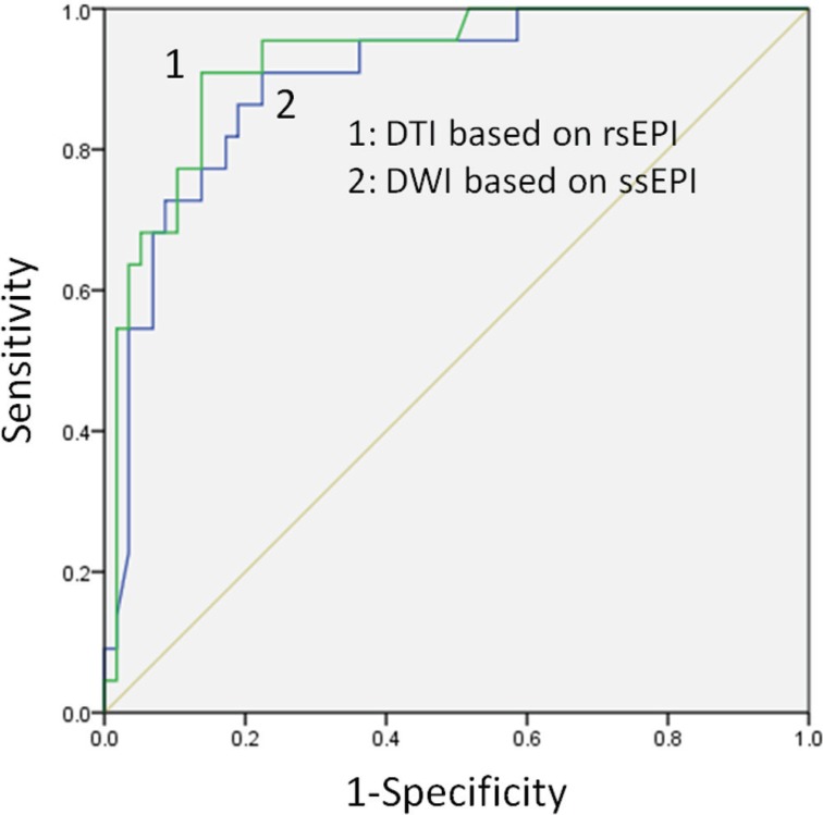
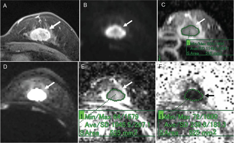
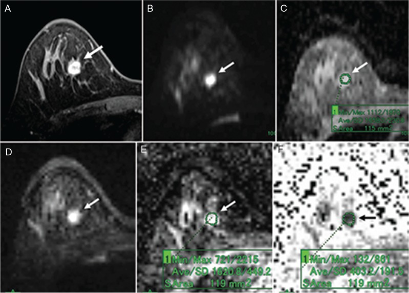
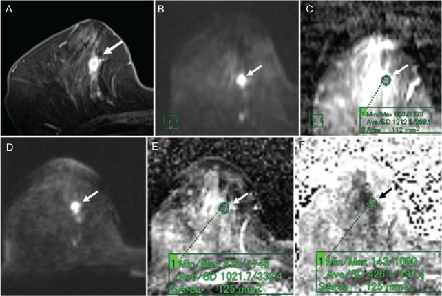
This retrospective study examined 80 pathologically proven breast lesions (22 benign and 58 malignant lesions) of 80 patients who underwent both diffusion-weighted imaging based on single-shot echo-planar imaging (DWI based on ss-EPI) and DTI based on rs-EPI with b-values of 0 and 1000. We identified and compared the diagnostic performances of the DWI based on ss-EPI and the DTI based on rs-EPI using ADCs by conducting a receiver-operating-characteristics (ROC) analysis. We determined the correlations between the ADCs and the prognostic markers and those of the FA values and the same markers.

RESULTS

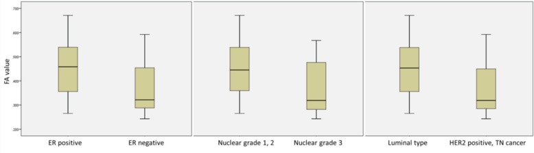
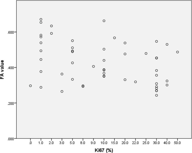
The median ADCs of the benign and malignant lesions based on the ss-EPI were 1.57 and 1.2 × 10 mm/sec, and those based on the rs-EPI were 1.53 and 1.09 × 10 mm/sec, respectively. The area under the curve on the ROC analysis based on rs-EPI (0.924) was greater than that based on ss-EPI (0.897). There were no significant correlations between the ADCs and the prognostic markers, but there were significant correlations between the FA values and the estrogen receptor status, a proliferative marker, the nuclear grade and the intrinsic subtype.

CONCLUSION

For breast cancer, DTI based on rs-EPI had superior diagnostic performance compared to DWI based on ss-EPI. Compared with the ADCs, the FA values were more closely correlated with prognostic markers of invasive breast cancer.

摘要

目的

评估读出分段回波平面扩散张量成像(基于读出分割回波平面成像的扩散张量成像,rs-EPI-DTI)对乳腺癌的诊断性能,并确定基于rs-EPI-DTI获得的表观扩散系数(ADC)和分数各向异性(FA)值与浸润性乳腺癌预后标志物之间的相关性。

材料与方法

本回顾性研究检查了80例患者的80个经病理证实的乳腺病变(22个良性病变和58个恶性病变),这些患者均接受了基于单次激发回波平面成像的扩散加权成像(基于单次激发回波平面成像的扩散加权成像,ss-EPI-DWI)和b值为0和1000的基于rs-EPI的扩散张量成像(rs-EPI-DTI)。我们通过进行受试者操作特征(ROC)分析,使用ADC来识别和比较基于ss-EPI的扩散加权成像和基于rs-EPI的扩散张量成像的诊断性能。我们确定了ADC与预后标志物之间的相关性以及FA值与相同标志物之间的相关性。

结果

基于ss-EPI的良性和恶性病变的ADC中位数分别为1.57和1.2×10⁻³mm²/sec,基于rs-EPI的分别为1.53和1.09×10⁻³mm²/sec。基于rs-EPI的ROC分析曲线下面积(0.924)大于基于ss-EPI的(0.897)。ADC与预后标志物之间无显著相关性,但FA值与雌激素受体状态、增殖标志物、核分级和内在亚型之间存在显著相关性。

结论

对于乳腺癌,基于rs-EPI的扩散张量成像比基于ss-EPI的扩散加权成像具有更好的诊断性能。与ADC相比,FA值与浸润性乳腺癌的预后标志物相关性更强。

https://cdn.ncbi.nlm.nih.gov/pmc/blobs/ab58/5600032/cf07cdf363a7/mrms-16-245-g6.jpg
https://cdn.ncbi.nlm.nih.gov/pmc/blobs/ab58/5600032/ca5d3828acd4/mrms-16-245-g1.jpg
https://cdn.ncbi.nlm.nih.gov/pmc/blobs/ab58/5600032/8543ccbd7f05/mrms-16-245-g2.jpg
https://cdn.ncbi.nlm.nih.gov/pmc/blobs/ab58/5600032/cdcbc0065d01/mrms-16-245-g3.jpg
https://cdn.ncbi.nlm.nih.gov/pmc/blobs/ab58/5600032/5a4794574d39/mrms-16-245-g4.jpg
https://cdn.ncbi.nlm.nih.gov/pmc/blobs/ab58/5600032/52be9fac9971/mrms-16-245-g5.jpg
https://cdn.ncbi.nlm.nih.gov/pmc/blobs/ab58/5600032/cf07cdf363a7/mrms-16-245-g6.jpg

相似文献

1
Diagnostic Performance of Diffusion Tensor Imaging with Readout-segmented Echo-planar Imaging for Invasive Breast Cancer: Correlation of ADC and FA with Pathological Prognostic Markers.读出分段回波平面成像的扩散张量成像对浸润性乳腺癌的诊断性能:表观扩散系数(ADC)和各向异性分数(FA)与病理预后标志物的相关性
Magn Reson Med Sci. 2017 Jul 10;16(3):245-252. doi: 10.2463/mrms.mp.2016-0037. Epub 2016 Nov 16.
2
Readout-segmented echo-planar diffusion-weighted imaging in the assessment of orbital tumors: comparison with conventional single-shot echo-planar imaging in image quality and diagnostic performance.读出分段回波平面扩散加权成像在眼眶肿瘤评估中的应用:与传统单次激发回波平面成像在图像质量和诊断性能方面的比较。
Acta Radiol. 2017 Dec;58(12):1457-1467. doi: 10.1177/0284185117695667. Epub 2017 Mar 22.
3
Bilateral diffusion-weighted MR imaging of breast tumors with submillimeter resolution using readout-segmented echo-planar imaging at 7 T.使用 7T 读出分段回波平面成像技术对乳腺肿瘤进行亚毫米分辨率的双侧扩散加权磁共振成像。
Radiology. 2015 Jan;274(1):74-84. doi: 10.1148/radiol.14132340. Epub 2014 Oct 23.
4
Diffusion-Weighted MRI to Assess Sacroiliitis: Improved Image Quality and Diagnostic Performance of Readout-Segmented Echo-Planar Imaging (EPI) Over Conventional Single-Shot EPI.扩散加权 MRI 评估中轴型脊柱关节炎:分段读出回波平面成像(EPI)优于传统单次激发 EPI,可提高图像质量和诊断性能。
AJR Am J Roentgenol. 2021 Aug;217(2):450-459. doi: 10.2214/AJR.20.23953. Epub 2021 Jun 9.
5
Diagnostic value of multi-model high-resolution diffusion-weighted MR imaging in breast lesions: Based on simultaneous multi-slice readout-segmented echo-planar imaging.多模态高分辨率弥散加权磁共振成像在乳腺病变中的诊断价值:基于同时多层分段读出回波平面成像。
Eur J Radiol. 2022 Sep;154:110439. doi: 10.1016/j.ejrad.2022.110439. Epub 2022 Jul 14.
6
Analysis of Apparent Diffusion Coefficients of the Brain in Healthy Controls: A Comparison Study between Single-Shot Echo-Planar Imaging and Read-out-Segmented Echo-Planar Imaging.健康对照者脑表观扩散系数分析:单次激发回波平面成像与分段读出回波平面成像的对比研究。
Korean J Radiol. 2019 Jul;20(7):1138-1145. doi: 10.3348/kjr.2018.0899.
7
Readout-Segmented Echo-Planar Diffusion-Weighted MR Imaging Improves the Differentiation of Breast Cancer Receptor Statuses Compared With Conventional Diffusion-Weighted Imaging.读出分段回波平面扩散加权磁共振成像与传统扩散加权成像相比,可提高乳腺癌受体状态的鉴别能力。
J Magn Reson Imaging. 2022 Sep;56(3):691-699. doi: 10.1002/jmri.28065. Epub 2022 Jan 17.
8
Readout-segmented echo-planar imaging improves the image quality of diffusion-weighted MR imaging in rectal cancer: Comparison with single-shot echo-planar diffusion-weighted sequences.读出分段回波平面成像改善直肠癌扩散加权磁共振成像的图像质量:与单次激发回波平面扩散加权序列的比较。
Eur J Radiol. 2016 Oct;85(10):1818-1823. doi: 10.1016/j.ejrad.2016.08.008. Epub 2016 Aug 10.
9
Readout-segmented echo-planar imaging in diffusion-weighted imaging of the kidney: comparison with single-shot echo-planar imaging in image quality.分段读出回波平面成像在肾脏扩散加权成像中的应用:与单次激发回波平面成像在图像质量方面的比较。
Abdom Radiol (NY). 2016 Jan;41(1):100-8. doi: 10.1007/s00261-015-0615-5.
10
Diffusion-Weighted Imaging-guided MR Spectroscopy in Breast Lesions using Readout-Segmented Echo-Planar Imaging.使用读出分段回波平面成像的扩散加权成像引导的乳腺病变磁共振波谱分析
Eur Radiol. 2016 Jun;26(6):1565-74. doi: 10.1007/s00330-015-4000-0. Epub 2015 Sep 19.

引用本文的文献

1
Diffusion tensor imaging: survival analysis prediction in breast cancer patients.弥散张量成像:乳腺癌患者的生存分析预测。
Radiologie (Heidelb). 2024 Nov;64(Suppl 1):54-59. doi: 10.1007/s00117-023-01254-0. Epub 2024 Jan 26.
2
Multiparametric Approach to Breast Cancer With Emphasis on Magnetic Resonance Imaging in the Era of Personalized Breast Cancer Treatment.多参数方法在乳腺癌诊治中的应用——个体化乳腺癌治疗时代的磁共振成像重点
Invest Radiol. 2024 Jan 1;59(1):26-37. doi: 10.1097/RLI.0000000000001044. Epub 2023 Nov 22.
3
High-resolution diffusion-weighted imaging compared with conventional diffusion-weighted imaging and dynamic contrast-enhanced magnetic resonance imaging with regard to image quality and assessment of breast cancer morphology.

本文引用的文献

1
Correlation between 3T apparent diffusion coefficient values and grading of invasive breast carcinoma.3T 表观扩散系数值与浸润性乳腺癌分级之间的相关性
Eur J Radiol. 2014 Dec;83(12):2144-2150. doi: 10.1016/j.ejrad.2014.09.015. Epub 2014 Sep 30.
2
Luminal-type breast cancer: correlation of apparent diffusion coefficients with the Ki-67 labeling index.腔面型乳腺癌:表观扩散系数与 Ki-67 标记指数的相关性。
Radiology. 2015 Jan;274(1):66-73. doi: 10.1148/radiol.14140283. Epub 2014 Sep 5.
3
Apparent diffusion coefficient on 3.0 Tesla magnetic resonance imaging and prognostic factors in breast cancer.
高分辨率弥散加权成像与常规弥散加权成像和动态对比增强磁共振成像在乳腺癌形态学评估方面的图像质量比较。
Diagn Interv Radiol. 2023 Mar 29;29(2):251-259. doi: 10.5152/dir.2022.21362. Epub 2023 Feb 10.
4
The road to breast cancer screening with diffusion MRI.通过扩散磁共振成像进行乳腺癌筛查之路。
Front Oncol. 2023 Feb 21;13:993540. doi: 10.3389/fonc.2023.993540. eCollection 2023.
5
Diffusion Breast MRI: Current Standard and Emerging Techniques.扩散加权乳腺磁共振成像:当前标准与新兴技术
Front Oncol. 2022 Jul 8;12:844790. doi: 10.3389/fonc.2022.844790. eCollection 2022.
6
Breast Cancer Subtypes and Quantitative Magnetic Resonance Imaging: A Systemic Review.乳腺癌亚型与定量磁共振成像:一项系统综述
Life (Basel). 2022 Mar 28;12(4):490. doi: 10.3390/life12040490.
7
Readout-Segmented Echo-Planar Diffusion-Weighted MR Imaging Improves the Differentiation of Breast Cancer Receptor Statuses Compared With Conventional Diffusion-Weighted Imaging.读出分段回波平面扩散加权磁共振成像与传统扩散加权成像相比,可提高乳腺癌受体状态的鉴别能力。
J Magn Reson Imaging. 2022 Sep;56(3):691-699. doi: 10.1002/jmri.28065. Epub 2022 Jan 17.
8
Diagnostic Value of Combined Intravoxel Incoherent Motion Diffusion-Weighted Magnetic Resonance Imaging with Diffusion Tensor Imaging in Predicting Parametrial Infiltration in Cervical Cancer.磁共振体素内不相干运动扩散加权成像联合扩散张量成像对预测宫颈癌宫旁浸润的诊断价值。
Contrast Media Mol Imaging. 2021 May 11;2021:6651070. doi: 10.1155/2021/6651070. eCollection 2021.
9
Improving the image quality of DWI in breast cancer: comparison of multi-shot DWI using multiplexed sensitivity encoding to conventional single-shot echo-planar imaging DWI.提高乳腺癌 DWI 图像质量:多重敏感编码多激发 DWI 与常规单次激发 echo-planar 成像 DWI 的比较。
Br J Radiol. 2021 Mar 1;94(1119):20200427. doi: 10.1259/bjr.20200427. Epub 2020 Sep 9.
10
Evaluation of Malignant Breast Lesions Using High-resolution Readout-segmented Diffusion-weighted Echo-planar Imaging: Comparison with Pathology.高分辨率分段读出扩散加权回波平面成像在乳腺良恶性病变评估中的应用:与病理对照。
Magn Reson Med Sci. 2021 Jun 1;20(2):204-215. doi: 10.2463/mrms.mp.2020-0021. Epub 2020 Jul 2.
3.0特斯拉磁共振成像的表观扩散系数与乳腺癌的预后因素
Eur J Gynaecol Oncol. 2014;35(4):408-14.
4
Diffusion magnetic resonance imaging in breast cancer characterisation: correlations between the apparent diffusion coefficient and major prognostic factors.扩散磁共振成像在乳腺癌特征描述中的应用:表观扩散系数与主要预后因素的相关性。
Radiol Med. 2015 Mar;120(3):268-76. doi: 10.1007/s11547-014-0442-8. Epub 2014 Aug 6.
5
Readout-segmented echo-planar imaging in diffusion-weighted mr imaging in breast cancer: comparison with single-shot echo-planar imaging in image quality.乳腺癌扩散加权磁共振成像中的读出分割回波平面成像:与单次激发回波平面成像的图像质量比较
Korean J Radiol. 2014 Jul-Aug;15(4):403-10. doi: 10.3348/kjr.2014.15.4.403. Epub 2014 Jul 9.
6
Diffusion-tensor MR imaging of the breast: hormonal regulation.弥散张量磁共振成像在乳腺中的应用:激素调节。
Radiology. 2014 Jun;271(3):672-80. doi: 10.1148/radiol.14132084. Epub 2014 Feb 12.
7
Correlations between apparent diffusion coefficient values of invasive ductal carcinoma and pathologic factors on diffusion-weighted MRI at 3.0 Tesla.3.0特斯拉磁共振扩散加权成像中浸润性导管癌表观扩散系数值与病理因素的相关性
J Magn Reson Imaging. 2015 Jan;41(1):175-82. doi: 10.1002/jmri.24519. Epub 2013 Dec 18.
8
Decision making for breast lesions initially detected at contrast-enhanced breast MRI.乳腺病变的磁共振成像对比增强检查初步诊断的决策。
AJR Am J Roentgenol. 2013 Dec;201(6):1376-85. doi: 10.2214/AJR.12.8953.
9
High-resolution diffusion-weighted imaging for the separation of benign from malignant BI-RADS 4/5 lesions found on breast MRI at 3T.3T乳腺MRI上发现的BI-RADS 4/5类病变中,利用高分辨率扩散加权成像鉴别良恶性
J Magn Reson Imaging. 2014 Sep;40(3):674-81. doi: 10.1002/jmri.24416. Epub 2013 Nov 8.
10
Optimization of the parameters for diffusion tensor magnetic resonance imaging data acquisition for breast fiber tractography at 1.5 T.1.5T 磁共振弥散张量成像数据采集参数优化用于乳腺纤维束示踪成像
Clin Breast Cancer. 2014 Feb;14(1):61-7. doi: 10.1016/j.clbc.2013.09.002. Epub 2013 Sep 27.