Hertig Gabriel, Zehnder Matthias, Woloszyk Anna, Mitsiadis Thimios A, Ivica Anja, Weber Franz E
Oral Biotechnology and Bioengineering, University of Zurich Zurich, Switzerland.
Preventive Dentistry, Periodontology, and Cariology, University of Zurich Zurich, Switzerland.
Front Physiol. 2017 Mar 15;8:152. doi: 10.3389/fphys.2017.00152. eCollection 2017.
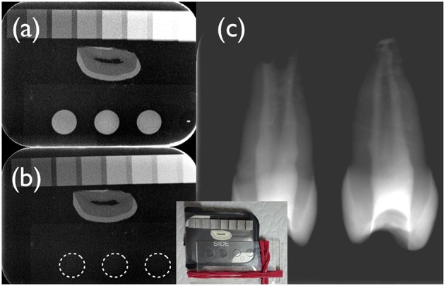
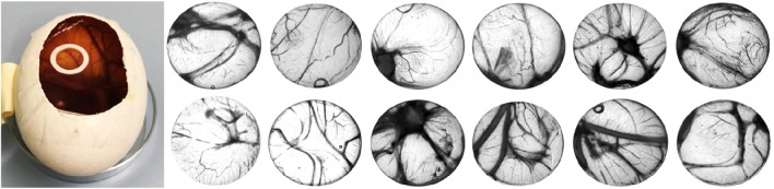
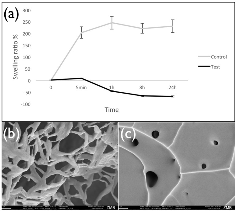
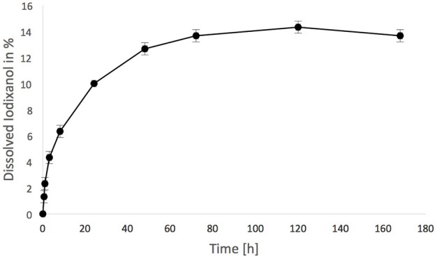
The application of biomaterials used in regenerative endodontics should be traceable. In this study, we checked some basic effects of rendering a fibrin hydrogel radiopaque using an iodine-based contrast agent (iodixanol) approved for systemic application. Fibrin hydrogels were prepared from a fibrin sealant (Tisseel) using either an isotonic iodixanol solution (Visipaque 320, test) or Tris buffer (control) as a diluent. Gelation kinetics, radiopacity, and swelling of lyophilized hydrogels were tested using standard methods. Hydrogel structure was evaluated using scanning electron microscopy (SEM). Furthermore, iodixanol release from the test gels was assessed using spectrophotometry, and tissue compatibility was compared between test and control hydrogels using the chick chorioallantoic membrane (CAM) assay. Results were compared using pairwise -test, < 0.05. Iodixanol caused a 70-fold delay in gelation to 26 min in the test compared to the control hydrogels (22 ± 1 s). Radiopacity of the test gels was 1.9 ± 0.2 mm Al/mm, compared to zero in the control hydrogels. Lyophilized hydrogel swelling was strongly reduced when iodixanol was added to the hydrogel ( < 0.05). Test hydrogels had an altered SEM appearance compared to controls, and exhibited a reduced porosity. Iodixanol release from the test hydrogels reached 14.5 ± 0.5% after 120 h and then ceased. This release did not have any apparent toxic effect and neither affected the viability, nor the physiology or vascularization of the CAM of fertilized chicken eggs. Iodixanol can render a fibrin hydrogel radiopaque and maintains its tissue compatibility, yet impacts gelation kinetics and hydrogel porosity.
用于再生牙髓病学的生物材料的应用应该是可追踪的。在本研究中,我们检测了使用一种经全身应用批准的碘基造影剂(碘克沙醇)使纤维蛋白水凝胶具有放射性不透明的一些基本效果。纤维蛋白水凝胶由纤维蛋白密封剂(Tisseel)制备,使用等渗碘克沙醇溶液(威视派克320,试验组)或Tris缓冲液(对照组)作为稀释剂。使用标准方法测试冻干水凝胶的凝胶化动力学、放射性不透明度和膨胀情况。使用扫描电子显微镜(SEM)评估水凝胶结构。此外,使用分光光度法评估试验凝胶中碘克沙醇的释放,并使用鸡胚绒毛尿囊膜(CAM)试验比较试验组和对照组水凝胶之间的组织相容性。使用配对检验比较结果,P<0.05。与对照水凝胶(22±1秒)相比,碘克沙醇使试验组的凝胶化延迟70倍至26分钟。试验凝胶的放射性不透明度为1.9±0.2毫米铝当量/毫米,而对照水凝胶为零。当向水凝胶中添加碘克沙醇时,冻干水凝胶的膨胀显著降低(P<0.05)。与对照组相比,试验水凝胶的SEM外观有所改变,且孔隙率降低。试验水凝胶中碘克沙醇的释放在120小时后达到14.5±0.5%,然后停止。这种释放没有任何明显的毒性作用,既不影响受精鸡蛋CAM的活力、生理学,也不影响其血管形成。碘克沙醇可使纤维蛋白水凝胶具有放射性不透明,并保持其组织相容性,但会影响凝胶化动力学和水凝胶孔隙率。