Department of Molecular and Systems Biology, Geisel School of Medicine at Dartmouth, Lebanon, NH, USA.
Department of Pediatrics, Geisel School of Medicine at Dartmouth, Lebanon, NH, USA.
Pediatr Res. 2017 Jul;82(1):164-172. doi: 10.1038/pr.2017.102. Epub 2017 May 3.
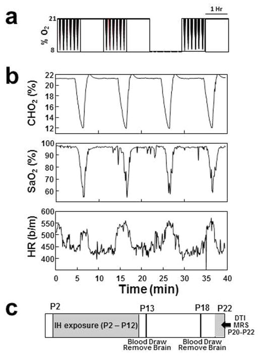
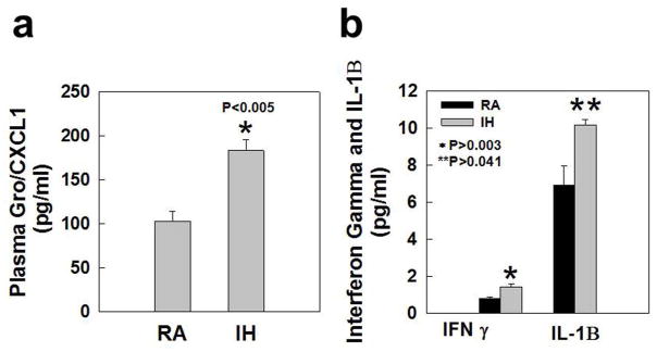
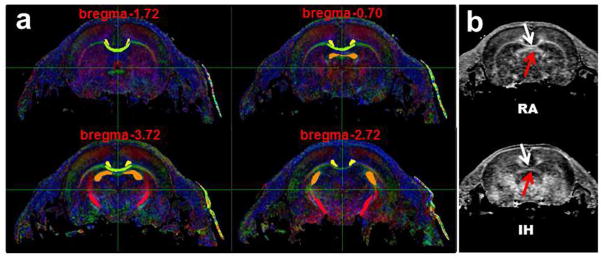
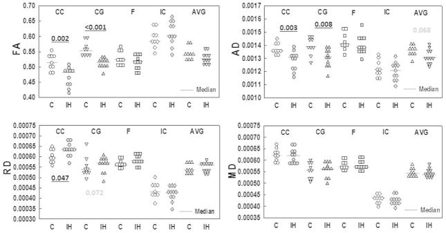
BackgroundPreterm infants are frequently exposed to intermittent hypoxia (IH) associated with apnea and periodic breathing that may result in inflammation and brain injury that later manifests as cognitive and executive function deficits. We used a rodent model to determine whether early postnatal exposure to IH would result in inflammation and brain injury.MethodsRat pups were exposed to IH from P2 to P12. Control animals were exposed to room air. Cytokines were analyzed in plasma and brain tissue at P13 and P18. At P20-P22, diffusion tensor imaging (DTI) and magnetic resonance spectroscopy (MRS) were performed.ResultsPups exposed to IH had increased plasma Gro/CXCL1 and cerebellar IFN-γ and IL-1β at P13, and brainstem enolase at P18. DTI showed a decrease in FA and AD in the corpus callosum (CC) and cingulate gyrus, and an increase in RD in the CC. MRS revealed decreases in NAA/Cho, Cr, Tau/Cr, and Gly/Cr; increases in TCho and GPC in the brainstem; and decreases in NAA/Cho in the hippocampus.ConclusionsWe conclude that early postnatal exposure to IH, similar in magnitude to that experienced in human preterm infants, is associated with evidence for proinflammatory changes, decreases in white matter integrity, and metabolic changes consistent with hypoxia.
早产儿经常会经历与呼吸暂停和周期性呼吸相关的间歇性低氧(IH),这可能导致炎症和脑损伤,进而表现为认知和执行功能缺陷。我们使用了一种啮齿动物模型来确定早期的 IH 暴露是否会导致炎症和脑损伤。
从 P2 到 P12,新生大鼠接受 IH 暴露。对照动物暴露在空气中。在 P13 和 P18 时分析了血浆和脑组织中的细胞因子。在 P20-P22 时,进行了弥散张量成像(DTI)和磁共振波谱(MRS)检查。
接受 IH 暴露的幼鼠在 P13 时血浆中的 Gro/CXCL1 和小脑 IFN-γ 和 IL-1β 增加,P18 时脑桥烯醇酶增加。DTI 显示胼胝体(CC)和扣带回的 FA 和 AD 降低,CC 的 RD 增加。MRS 显示脑桥中 NAA/Cho、Cr、Tau/Cr 和 Gly/Cr 减少,TCho 和 GPC 增加,海马中 NAA/Cho 减少。
我们的结论是,与人类早产儿经历的 IH 相似的早期产后暴露与促炎变化、白质完整性下降以及与缺氧一致的代谢变化有关。