• 文献检索
  • 文档翻译
  • 深度研究
  • 学术资讯
  • Suppr Zotero 插件Zotero 插件
  • 邀请有礼
  • 套餐&价格
  • 历史记录
应用&插件
Suppr Zotero 插件Zotero 插件浏览器插件Mac 客户端Windows 客户端微信小程序
定价
高级版会员购买积分包购买API积分包
服务
文献检索文档翻译深度研究API 文档MCP 服务
关于我们
关于 Suppr公司介绍联系我们用户协议隐私条款
关注我们

Suppr 超能文献

核心技术专利:CN118964589B侵权必究
粤ICP备2023148730 号-1Suppr @ 2026

文献检索

告别复杂PubMed语法,用中文像聊天一样搜索,搜遍4000万医学文献。AI智能推荐,让科研检索更轻松。

立即免费搜索

文件翻译

保留排版,准确专业,支持PDF/Word/PPT等文件格式,支持 12+语言互译。

免费翻译文档

深度研究

AI帮你快速写综述,25分钟生成高质量综述,智能提取关键信息,辅助科研写作。

立即免费体验

扩散加权磁共振成像可在神经肿胀的情况下对早期轴突损失进行量化。

Diffusion MRI quantifies early axonal loss in the presence of nerve swelling.

作者信息

Lin Tsen-Hsuan, Chiang Chia-Wen, Perez-Torres Carlos J, Sun Peng, Wallendorf Michael, Schmidt Robert E, Cross Anne H, Song Sheng-Kwei

机构信息

Radiology, Washington University School of Medicine, 660 S Euclid Ave, St. Louis, MO, 63110, USA.

Current Address: Institute of Biomedical Engineering and Nanomedicine, National Health Research Institute, 35 Keyan Road, Zhunan, Miaoli County, 35053, Taiwan.

出版信息

J Neuroinflammation. 2017 Apr 7;14(1):78. doi: 10.1186/s12974-017-0852-3.

DOI:10.1186/s12974-017-0852-3
PMID:28388913
原文链接:https://pmc.ncbi.nlm.nih.gov/articles/PMC5384143/
Abstract

BACKGROUND

Magnetic resonance imaging markers have been widely used to detect and quantify white matter pathologies in multiple sclerosis. We have recently developed a diffusion basis spectrum imaging (DBSI) to distinguish and quantify co-existing axonal injury, demyelination, and inflammation in multiple sclerosis patients and animal models. It could serve as a longitudinal marker for axonal loss, a primary cause of permanent neurological impairments and disease progression.

METHODS

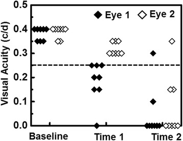
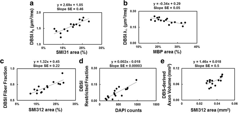
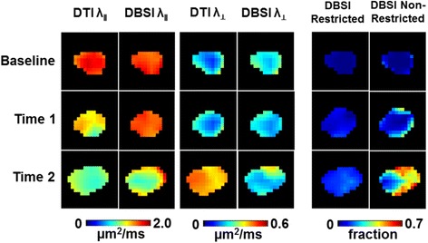
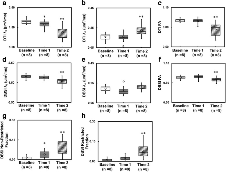
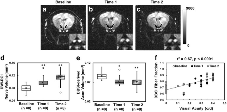
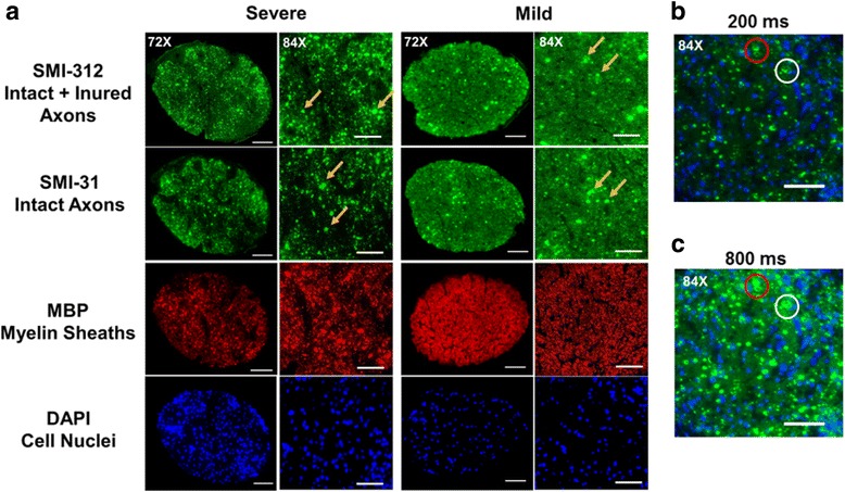
Eight 10-week-old female C57BL/6 mice underwent optic nerve DBSI, followed by a week-long recuperation prior to active immunization for experimental autoimmune encephalomyelitis (EAE). Visual acuity of all mice was assessed daily. Longitudinal DBSI was performed in mouse optic nerves at baseline (naïve, before immunization), before, during, and after the onset of optic neuritis. Tissues were perfusion fixed after final in vivo scans. The correlation between DBSI detected pathologies and corresponding immunohistochemistry markers was quantitatively assessed.

RESULTS

In this cohort of EAE mice, monocular vision impairment occurred in all animals. In vivo DBSI detected, differentiated, and quantified optic nerve inflammation, demyelination, and axonal injury/loss, correlating nerve pathologies with visual acuity at different time points of acute optic neuritis. DBSI quantified, in the presence of optic nerve swelling, ~15% axonal loss at the onset of optic neuritis in EAE mice.

CONCLUSIONS

Our findings support the notion that axonal loss could occur early in EAE mice. DBSI detected pathologies in the posterior visual pathway unreachable by optical coherence tomography and without confounding inflammation induced optic nerve swelling. DBSI could thus decipher the interrelationship among various pathological components and the role each plays in disease progression. Quantification of the rate of axonal loss could potentially serve as the biomarker to predict treatment outcome and to determine when progressive disease starts.

摘要

背景

磁共振成像标记物已被广泛用于检测和量化多发性硬化症中的白质病变。我们最近开发了一种扩散基谱成像(DBSI)技术,用于区分和量化多发性硬化症患者及动物模型中并存的轴突损伤、脱髓鞘和炎症。它可作为轴突丢失的纵向标记物,轴突丢失是永久性神经功能障碍和疾病进展的主要原因。

方法

八只10周龄雌性C57BL/6小鼠接受视神经DBSI检查,随后在主动免疫诱导实验性自身免疫性脑脊髓炎(EAE)前进行为期一周的恢复。每天评估所有小鼠的视力。在基线(未免疫、免疫前)、视神经炎发作前、发作期间和发作后,对小鼠视神经进行纵向DBSI检查。在最后一次体内扫描后对组织进行灌注固定。定量评估DBSI检测到的病变与相应免疫组化标记物之间的相关性。

结果

在这组EAE小鼠中,所有动物均出现单眼视力损害。体内DBSI检测、区分并量化了视神经炎症、脱髓鞘和轴突损伤/丢失,将神经病变与急性视神经炎不同时间点的视力相关联。在视神经肿胀的情况下,DBSI量化了EAE小鼠视神经炎发作时约15%的轴突丢失。

结论

我们的研究结果支持轴突丢失可能在EAE小鼠早期发生的观点。DBSI检测到光学相干断层扫描无法到达的后视觉通路中的病变,且不受炎症引起的视神经肿胀的干扰。因此,DBSI可以解读各种病理成分之间的相互关系以及它们在疾病进展中各自所起的作用。轴突丢失率的量化可能作为预测治疗结果和确定疾病何时开始进展的生物标志物。

https://cdn.ncbi.nlm.nih.gov/pmc/blobs/b940/5384143/f4231c91de9e/12974_2017_852_Fig6_HTML.jpg
https://cdn.ncbi.nlm.nih.gov/pmc/blobs/b940/5384143/d99d802452c8/12974_2017_852_Fig1_HTML.jpg
https://cdn.ncbi.nlm.nih.gov/pmc/blobs/b940/5384143/15f26d1d89be/12974_2017_852_Fig2_HTML.jpg
https://cdn.ncbi.nlm.nih.gov/pmc/blobs/b940/5384143/db06ef6aa9f1/12974_2017_852_Fig3_HTML.jpg
https://cdn.ncbi.nlm.nih.gov/pmc/blobs/b940/5384143/3a25b0452e69/12974_2017_852_Fig4_HTML.jpg
https://cdn.ncbi.nlm.nih.gov/pmc/blobs/b940/5384143/b5bcd67af44c/12974_2017_852_Fig5_HTML.jpg
https://cdn.ncbi.nlm.nih.gov/pmc/blobs/b940/5384143/f4231c91de9e/12974_2017_852_Fig6_HTML.jpg

相似文献

1
Diffusion MRI quantifies early axonal loss in the presence of nerve swelling.扩散加权磁共振成像可在神经肿胀的情况下对早期轴突损失进行量化。
J Neuroinflammation. 2017 Apr 7;14(1):78. doi: 10.1186/s12974-017-0852-3.
2
Diffusion basis spectrum imaging measures anti-inflammatory and neuroprotective effects of fingolimod on murine optic neuritis.扩散张量谱成像测量芬戈莫德对实验性变态反应性视神经炎的抗炎和神经保护作用。
Neuroimage Clin. 2021;31:102732. doi: 10.1016/j.nicl.2021.102732. Epub 2021 Jun 17.
3
Exploring experimental autoimmune optic neuritis using multimodal imaging.探索实验性自身免疫性视神经炎的多模态成像。
Neuroimage. 2018 Jul 15;175:327-339. doi: 10.1016/j.neuroimage.2018.04.004. Epub 2018 Apr 6.
4
Diffusion fMRI detects white-matter dysfunction in mice with acute optic neuritis.扩散加权功能磁共振成像检测急性视神经炎小鼠的白质功能障碍。
Neurobiol Dis. 2014 Jul;67:1-8. doi: 10.1016/j.nbd.2014.02.007. Epub 2014 Mar 13.
5
Quantifying white matter tract diffusion parameters in the presence of increased extra-fiber cellularity and vasogenic edema.在存在额外纤维细胞增多和血管源性水肿的情况下量化白质束扩散参数。
Neuroimage. 2014 Nov 1;101:310-9. doi: 10.1016/j.neuroimage.2014.06.064. Epub 2014 Jul 11.
6
Diffusion Basis Spectrum Imaging Detects Axonal Loss After Transient Dexamethasone Treatment in Optic Neuritis Mice.扩散基础光谱成像检测视神经炎小鼠短暂地塞米松治疗后的轴突损失。
Front Neurosci. 2021 Jan 22;14:592063. doi: 10.3389/fnins.2020.592063. eCollection 2020.
7
Loss of Nrf2 exacerbates the visual deficits and optic neuritis elicited by experimental autoimmune encephalomyelitis.Nrf2的缺失会加剧实验性自身免疫性脑脊髓炎引发的视觉缺陷和视神经炎。
Mol Vis. 2016 Dec 30;22:1503-1513. eCollection 2016.
8
HE3286 reduces axonal loss and preserves retinal ganglion cell function in experimental optic neuritis.HE3286 减少实验性视神经炎中的轴突丢失并维持视网膜神经节细胞功能。
Invest Ophthalmol Vis Sci. 2014 Aug 19;55(9):5744-51. doi: 10.1167/iovs.14-14672.
9
Noninvasive Quantification of Axonal Loss in the Presence of Tissue Swelling in Traumatic Spinal Cord Injury Mice.在创伤性脊髓损伤小鼠存在组织肿胀的情况下,对轴突丢失进行无创定量。
J Neurotrauma. 2019 Aug 1;36(15):2308-2315. doi: 10.1089/neu.2018.6016. Epub 2019 Jan 11.
10
Inflammatory demyelination induces axonal injury and retinal ganglion cell apoptosis in experimental optic neuritis.在实验性视神经炎中,炎性脱髓鞘会导致轴突损伤和视网膜神经节细胞凋亡。
Exp Eye Res. 2008 Sep;87(3):208-13. doi: 10.1016/j.exer.2008.05.017. Epub 2008 Jun 3.

引用本文的文献

1
Relationship between sodium and diffusion MRI metrics in multiple sclerosis.多发性硬化症中钠与扩散磁共振成像指标之间的关系。
Brain Commun. 2025 Jan 6;7(1):fcae446. doi: 10.1093/braincomms/fcae446. eCollection 2025.
2
Diffusion basis spectrum imaging detects subclinical traumatic optic neuropathy in a closed-head impact mouse model of traumatic brain injury.扩散基谱成像在创伤性脑损伤的闭合性头部撞击小鼠模型中检测亚临床创伤性视神经病变。
Front Neurol. 2023 Dec 13;14:1269817. doi: 10.3389/fneur.2023.1269817. eCollection 2023.
3
Diffusion basis spectrum imaging detects pathological alterations in substantia nigra and white matter tracts with early-stage Parkinson's disease.

本文引用的文献

1
Differentiation and quantification of inflammation, demyelination and axon injury or loss in multiple sclerosis.多发性硬化症中炎症、脱髓鞘以及轴突损伤或丧失的鉴别与定量分析
Brain. 2015 May;138(Pt 5):1223-38. doi: 10.1093/brain/awv046. Epub 2015 Feb 26.
2
Vision and vision-related outcome measures in multiple sclerosis.多发性硬化症中的视力及与视力相关的预后指标
Brain. 2015 Jan;138(Pt 1):11-27. doi: 10.1093/brain/awu335. Epub 2014 Nov 28.
3
Treatment of acute optic neuritis and vision complaints in multiple sclerosis.多发性硬化症中急性视神经炎及视力问题的治疗
扩散张量谱成像检测早期帕金森病患者黑质和白质束的病理改变。
Eur Radiol. 2023 Dec;33(12):9109-9119. doi: 10.1007/s00330-023-09780-0. Epub 2023 Jul 12.
4
Diffusion basis spectrum imaging as an adjunct to conventional MRI leads to earlier diagnosis of high-grade glioma tumor progression versus treatment effect.扩散基波谱成像作为传统磁共振成像的辅助手段,相较于治疗效果,能更早地诊断高级别胶质瘤的肿瘤进展。
Neurooncol Adv. 2023 Apr 19;5(1):vdad050. doi: 10.1093/noajnl/vdad050. eCollection 2023 Jan-Dec.
5
Childhood obesity is linked to putative neuroinflammation in brain white matter, hypothalamus, and striatum.儿童肥胖与脑白质、下丘脑和纹状体中假定的神经炎症有关。
Cereb Cortex Commun. 2023 May 2;4(2):tgad007. doi: 10.1093/texcom/tgad007. eCollection 2023.
6
Neuroinflammation and white matter alterations in occupational manganese exposure assessed by diffusion basis spectrum imaging.职业性锰暴露的神经炎症和白质改变的扩散张量频谱成像评估。
Neurotoxicology. 2023 Jul;97:25-33. doi: 10.1016/j.neuro.2023.04.013. Epub 2023 Apr 29.
7
Magnetic resonance imaging of the monkey fetal brain in utero.猕猴胎儿子宫内脑部的磁共振成像
Investig Magn Reson Imaging. 2022 Dec;26(4):177-190. doi: 10.13104/imri.2022.26.4.177. Epub 2022 Dec 31.
8
3-Dimensional Immunostaining and Automated Deep-Learning Based Analysis of Nerve Degeneration.三维免疫染色和基于自动化深度学习的神经退行性变分析。
Int J Mol Sci. 2022 Nov 26;23(23):14811. doi: 10.3390/ijms232314811.
9
Characterization of multiple sclerosis neuroinflammation and neurodegeneration with relaxation and diffusion basis spectrum imaging.应用弛豫和弥散基谱成像技术对多发性硬化的神经炎症和神经退行性变进行特征描述。
Mult Scler. 2022 Mar;28(3):418-428. doi: 10.1177/13524585211023345. Epub 2021 Jun 16.
10
Diffusion Basis Spectrum Imaging Detects Axonal Loss After Transient Dexamethasone Treatment in Optic Neuritis Mice.扩散基础光谱成像检测视神经炎小鼠短暂地塞米松治疗后的轴突损失。
Front Neurosci. 2021 Jan 22;14:592063. doi: 10.3389/fnins.2020.592063. eCollection 2020.
Curr Treat Options Neurol. 2015 Jan;17(1):328. doi: 10.1007/s11940-014-0328-z.
4
Clinical trials to clinical use: using vision as a model for multiple sclerosis and beyond.从临床试验到临床应用:以视觉为例探讨多发性硬化症及其他疾病
J Neuroophthalmol. 2014 Sep;34 Suppl:S18-23. doi: 10.1097/WNO.0000000000000163.
5
Multiple sclerosis: current knowledge and future outlook.多发性硬化症:当前认知与未来展望。
Eur Neurol. 2014;72(3-4):132-41. doi: 10.1159/000360528. Epub 2014 Jul 30.
6
Phase-aligned multiple spin-echo averaging: a simple way to improve signal-to-noise ratio of in vivo mouse spinal cord diffusion tensor image.相位对齐的多次自旋回波平均法:一种提高体内小鼠脊髓扩散张量图像信噪比的简单方法。
Magn Reson Imaging. 2014 Dec;32(10):1335-43. doi: 10.1016/j.mri.2014.07.004. Epub 2014 Aug 1.
7
Quantifying white matter tract diffusion parameters in the presence of increased extra-fiber cellularity and vasogenic edema.在存在额外纤维细胞增多和血管源性水肿的情况下量化白质束扩散参数。
Neuroimage. 2014 Nov 1;101:310-9. doi: 10.1016/j.neuroimage.2014.06.064. Epub 2014 Jul 11.
8
Axonal transport rate decreased at the onset of optic neuritis in EAE mice.在实验性自身免疫性脑脊髓炎(EAE)小鼠中,视神经炎发作时轴突运输速率降低。
Neuroimage. 2014 Oct 15;100:244-53. doi: 10.1016/j.neuroimage.2014.06.009. Epub 2014 Jun 14.
9
Diffusion basis spectrum imaging detects and distinguishes coexisting subclinical inflammation, demyelination and axonal injury in experimental autoimmune encephalomyelitis mice.弥散张量谱成像可检测和区分实验性自身免疫性脑脊髓炎小鼠共存的亚临床炎症、脱髓鞘和轴突损伤。
NMR Biomed. 2014 Jul;27(7):843-52. doi: 10.1002/nbm.3129. Epub 2014 May 12.
10
Imaging outcomes for trials of remyelination in multiple sclerosis.多发性硬化症中髓鞘再生试验的成像结果。
J Neurol Neurosurg Psychiatry. 2014 Dec;85(12):1396-404. doi: 10.1136/jnnp-2014-307650. Epub 2014 Apr 25.