Zhao Ting, Zhao Chenyan, Zhou Yanting, Zheng Jing, Gao Shujun, Lu Yuan
Department of Gynecology, Obstetrics and Gynecology Hospital of Fudan University, Shanghai, China.
Department of Pathology, Obstetrics and Gynecology Hospital of Fudan University, Shanghai, China.
Cancer Med. 2017 May;6(5):1072-1081. doi: 10.1002/cam4.1053. Epub 2017 Apr 12.
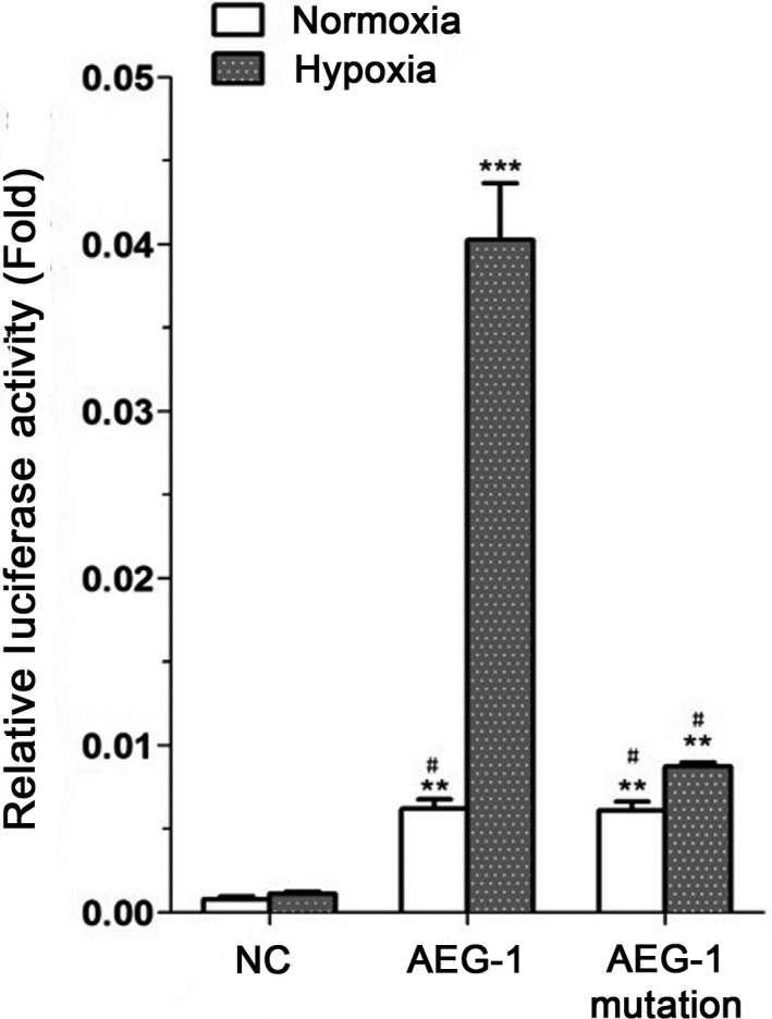
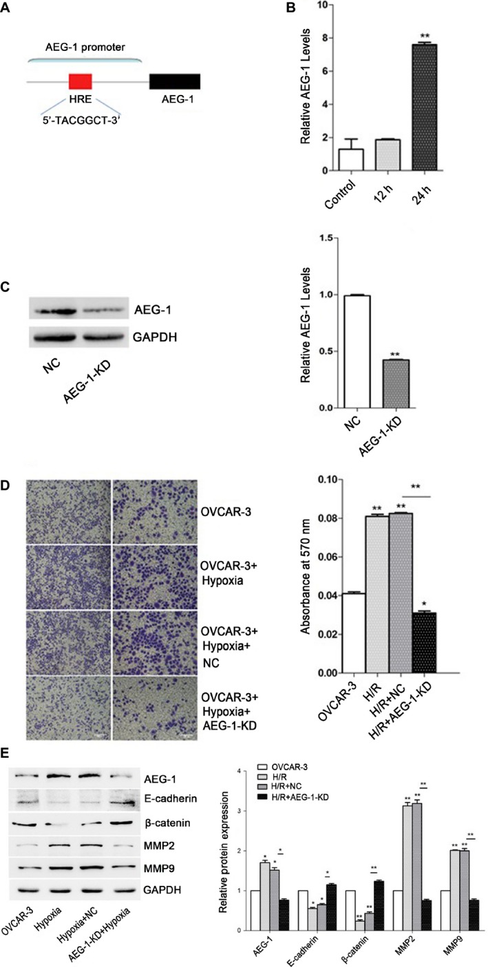
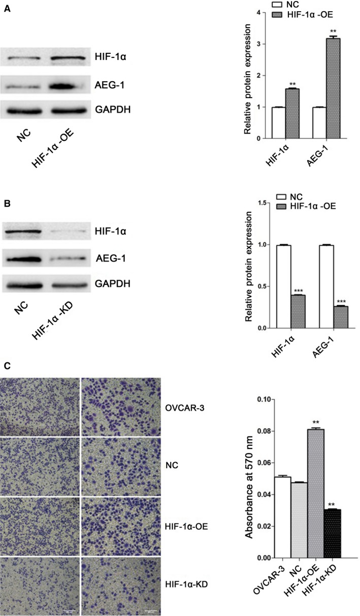
Ovarian cancer with the highest mortality rate among gynecological malignancies is one of common cancers among female cancer patients. As reported in recent years, AEG-1 was associated with the occurrence, development, and metastasis of ovarian cancer, but the mechanisms remain unclear. In the current study, invasion capabilities of ovarian cancer OVCAR3 cells were measured by viral infection and transwell assay. Western blot analysis was used to evaluate the expression levels of β-catenin, E-cadherin, MMP2, and MMP9. With qRT-PCR analysis, AEG-1 and HIF-1α gene expression were detected. We used luciferase reporter gene to measure AEG-1 promoter activity under normoxia/hypoxia in OVCAR3 cells. Our work demonstrated that AEG-1 significantly enhanced invasion capabilities of OVCAR3 cells and the expression levels of β-catenin, E-cadherin, MMP2, and MMP9 associated with invasion capabilities of OVCAR3 cells were upregulated. Furthermore, hypoxia enhanced invasion capabilities of OVCAR3 cells and induced AEG-1 high gene expression, which was reversed by AEG-1 knockdown lentivirus. HIF-1α expression upregulation was induced in OVCAR3 cells after hypoxia. HIF-1α knockdown lentivirus induced downregulated expression of AEG-1 and invasion capabilities of OVCAR3 cells were also inhibited. Wild-type AEG-1 promoter activity under hypoxic conditions was significantly higher than that AEG-1 mutation under normoxic conditions in the absence of hypoxia response. Our results suggested that HIF-1α binds to AEG-1 promoter to upregulate its expression, which was correlated with metastasis in ovarian cancer by inducing the expression of MMP2 and MMP9 as well as inhibiting expression of E-cadherin and β-catenin.
卵巢癌是妇科恶性肿瘤中死亡率最高的疾病之一,也是女性癌症患者中常见的癌症之一。近年来有报道称,AEG-1与卵巢癌的发生、发展和转移有关,但其机制仍不清楚。在本研究中,通过病毒感染和Transwell实验检测卵巢癌OVCAR3细胞的侵袭能力。采用蛋白质免疫印迹分析评估β-连环蛋白、E-钙黏蛋白、基质金属蛋白酶2(MMP2)和基质金属蛋白酶9(MMP9)的表达水平。通过qRT-PCR分析检测AEG-1和低氧诱导因子-1α(HIF-1α)基因表达。我们使用荧光素酶报告基因检测OVCAR3细胞在常氧/低氧条件下AEG-1启动子活性。我们的研究表明,AEG-1显著增强了OVCAR3细胞的侵袭能力,且与OVCAR3细胞侵袭能力相关的β-连环蛋白、E-钙黏蛋白、MMP2和MMP9的表达水平上调。此外,低氧增强了OVCAR3细胞的侵袭能力并诱导AEG-1高表达,而AEG-1敲低慢病毒可逆转这种情况。低氧后OVCAR3细胞中HIF-1α表达上调。HIF-1α敲低慢病毒诱导AEG-1表达下调,同时OVCAR3细胞的侵袭能力也受到抑制。在无低氧反应的情况下,低氧条件下野生型AEG-1启动子活性显著高于常氧条件下AEG-1突变体。我们的结果表明,HIF-1α与AEG-1启动子结合上调其表达,这通过诱导MMP2和MMP9的表达以及抑制E-钙黏蛋白和β-连环蛋白的表达与卵巢癌转移相关。