• 文献检索
  • 文档翻译
  • 深度研究
  • 学术资讯
  • Suppr Zotero 插件Zotero 插件
  • 邀请有礼
  • 套餐&价格
  • 历史记录
应用&插件
Suppr Zotero 插件Zotero 插件浏览器插件Mac 客户端Windows 客户端微信小程序
定价
高级版会员购买积分包购买API积分包
服务
文献检索文档翻译深度研究API 文档MCP 服务
关于我们
关于 Suppr公司介绍联系我们用户协议隐私条款
关注我们

Suppr 超能文献

核心技术专利:CN118964589B侵权必究
粤ICP备2023148730 号-1Suppr @ 2026

文献检索

告别复杂PubMed语法,用中文像聊天一样搜索,搜遍4000万医学文献。AI智能推荐,让科研检索更轻松。

立即免费搜索

文件翻译

保留排版,准确专业,支持PDF/Word/PPT等文件格式,支持 12+语言互译。

免费翻译文档

深度研究

AI帮你快速写综述,25分钟生成高质量综述,智能提取关键信息,辅助科研写作。

立即免费体验

无标记容积光学成像完整的小鼠大脑。

Label-free volumetric optical imaging of intact murine brains.

机构信息

Wellman Center for Photomedicine, Massachusetts General Hospital, Boston, MA 02114, USA.

Harvard Medical School, Boston, MA 02115, USA.

出版信息

Sci Rep. 2017 Apr 12;7:46306. doi: 10.1038/srep46306.

DOI:10.1038/srep46306
PMID:28401897
原文链接:https://pmc.ncbi.nlm.nih.gov/articles/PMC5388920/
Abstract

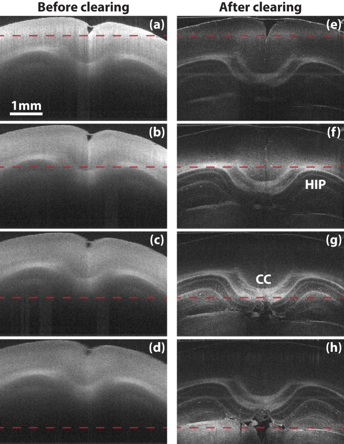
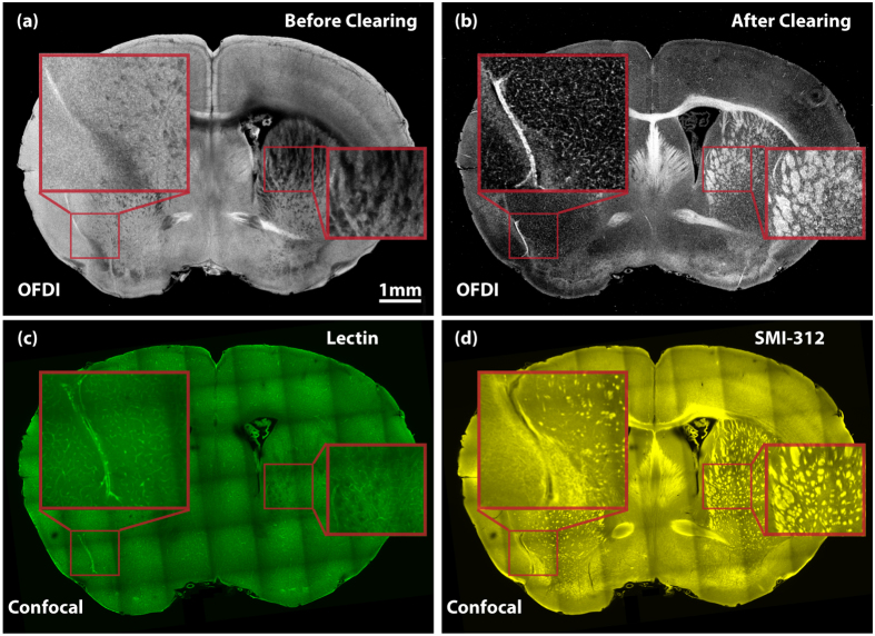
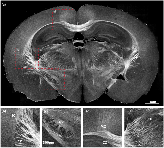
A central effort of today's neuroscience is to study the brain's 'wiring diagram'. The nervous system is believed to be a network of neurons interacting with each other through synaptic connection between axons and dendrites, therefore the neuronal connectivity map not only depicts the underlying anatomy, but also has important behavioral implications. Different approaches have been utilized to decipher neuronal circuits, including electron microscopy (EM) and light microscopy (LM). However, these approaches typically demand extensive sectioning and reconstruction for a brain sample. Recently, tissue clearing methods have enabled the investigation of a fully assembled biological system with greatly improved light penetration. Yet, most of these implementations, still require either genetic or exogenous contrast labeling for light microscopy. Here we demonstrate a high-speed approach, termed as Clearing Assisted Scattering Tomography (CAST), where intact brains can be imaged at optical resolution without labeling by leveraging tissue clearing and the scattering contrast of optical frequency domain imaging (OFDI).

摘要

当今神经科学的一个主要研究方向是研究大脑的“连接图”。人们认为神经系统是一个通过轴突和树突之间的突触连接相互作用的神经元网络,因此神经元连接图不仅描绘了潜在的解剖结构,而且具有重要的行为意义。已经有不同的方法被用于破译神经元回路,包括电子显微镜(EM)和光学显微镜(LM)。然而,这些方法通常需要对脑组织样本进行广泛的切片和重建。最近,组织透明化方法使得对完全组装的生物系统进行研究成为可能,其光穿透能力得到了极大的提高。然而,这些实现方法中的大多数仍然需要遗传或外源性对比标记来进行光学显微镜检测。在这里,我们展示了一种高速方法,称为 Clearing Assisted Scattering Tomography(CAST),它利用组织透明化和光频域成像(OFDI)的散射对比度,无需标记即可在不降低分辨率的情况下对完整的大脑进行成像。

https://cdn.ncbi.nlm.nih.gov/pmc/blobs/3241/5388920/ca8c4de32881/srep46306-f4.jpg
https://cdn.ncbi.nlm.nih.gov/pmc/blobs/3241/5388920/6ab02a89c258/srep46306-f1.jpg
https://cdn.ncbi.nlm.nih.gov/pmc/blobs/3241/5388920/7c027d26389e/srep46306-f2.jpg
https://cdn.ncbi.nlm.nih.gov/pmc/blobs/3241/5388920/fbf4f567a56d/srep46306-f3.jpg
https://cdn.ncbi.nlm.nih.gov/pmc/blobs/3241/5388920/ca8c4de32881/srep46306-f4.jpg

相似文献

1
Label-free volumetric optical imaging of intact murine brains.无标记容积光学成像完整的小鼠大脑。
Sci Rep. 2017 Apr 12;7:46306. doi: 10.1038/srep46306.
2
Whole Murine Brain Imaging Based on Optical Elastic Scattering.基于光弹性散射的全鼠脑成像。
Adv Exp Med Biol. 2021;3233:109-125. doi: 10.1007/978-981-15-7627-0_6.
3
UbasM: An effective balanced optical clearing method for intact biomedical imaging.UbasM:一种用于完整生物医学成像的有效平衡光透明方法。
Sci Rep. 2017 Sep 22;7(1):12218. doi: 10.1038/s41598-017-12484-3.
4
Light sheet fluorescence microscopy for neuroscience.光片荧光显微镜在神经科学中的应用。
J Neurosci Methods. 2019 May 1;319:16-27. doi: 10.1016/j.jneumeth.2018.07.011. Epub 2018 Jul 23.
5
Can Developments in Tissue Optical Clearing Aid Super-Resolution Microscopy Imaging?组织光学透明化的发展能否辅助超分辨率显微镜成像?
Int J Mol Sci. 2021 Jun 23;22(13):6730. doi: 10.3390/ijms22136730.
6
Tissue clearing for optical anatomy.组织透明化用于光学解剖。
Angew Chem Int Ed Engl. 2013 Oct 11;52(42):10949-51. doi: 10.1002/anie.201306039. Epub 2013 Sep 3.
7
Optical Clearing and Labeling for Light-sheet Fluorescence Microscopy in Large-scale Human Brain Imaging.用于大尺度人脑成像的光片荧光显微镜的光透明和标记。
J Vis Exp. 2024 Jan 26(203). doi: 10.3791/65960.
8
SeeDB: a simple and morphology-preserving optical clearing agent for neuronal circuit reconstruction.SeeDB:一种简单且保持形态的光透明剂,用于神经元回路重建。
Nat Neurosci. 2013 Aug;16(8):1154-61. doi: 10.1038/nn.3447. Epub 2013 Jun 23.
9
Automatic and adaptive heterogeneous refractive index compensation for light-sheet microscopy.用于光片显微镜的自动自适应异质折射率补偿
Nat Commun. 2017 Sep 20;8(1):612. doi: 10.1038/s41467-017-00514-7.
10
FluoEM, virtual labeling of axons in three-dimensional electron microscopy data for long-range connectomics.FluoEM,用于三维电子显微镜数据中远距离连接组学的轴突虚拟标记。
Elife. 2018 Aug 14;7:e38976. doi: 10.7554/eLife.38976.

引用本文的文献

1
From superior contrast to super resolution label free optical microscopy.从高对比度到超分辨率无标记光学显微镜。
Npj Imaging. 2025 Jan 13;3(1):1. doi: 10.1038/s44303-024-00064-w.
2
Glass-brain mapping provides an adjunct tool for structural analysis in mouse models of neurodevelopmental disease.脑图谱为神经发育疾病小鼠模型的结构分析提供了一种辅助工具。
Neuroimage Rep. 2021 Jun 10;1(2):100023. doi: 10.1016/j.ynirp.2021.100023. eCollection 2021 Jun.
3
Probabilistic volumetric speckle suppression in OCT using deep learning.基于深度学习的光学相干断层扫描中的概率体积散斑抑制

本文引用的文献

1
Shrinkage-mediated imaging of entire organs and organisms using uDISCO.uDISCO 介导的整个器官和生物体的缩时成像。
Nat Methods. 2016 Oct;13(10):859-67. doi: 10.1038/nmeth.3964. Epub 2016 Aug 22.
2
Simple, Scalable Proteomic Imaging for High-Dimensional Profiling of Intact Systems.用于完整系统高维分析的简单、可扩展蛋白质组学成像
Cell. 2015 Dec 3;163(6):1500-14. doi: 10.1016/j.cell.2015.11.025.
3
Stochastic electrotransport selectively enhances the transport of highly electromobile molecules.随机电迁移选择性地增强了高电迁移性分子的迁移。
Biomed Opt Express. 2024 Jul 3;15(8):4453-4469. doi: 10.1364/BOE.523716. eCollection 2024 Aug 1.
4
Probabilistic volumetric speckle suppression in OCT using deep learning.基于深度学习的光学相干断层扫描中概率性体素散斑抑制
ArXiv. 2023 Dec 7:arXiv:2312.04460v1.
5
Endoscopic imaging of white matter fiber tracts using polarization-sensitive optical coherence tomography.应用偏振敏感光学相干断层成像术观察脑白质纤维束
Neuroimage. 2022 Dec 1;264:119755. doi: 10.1016/j.neuroimage.2022.119755. Epub 2022 Nov 15.
6
Multidimensional Imaging of Breast Cancer.乳腺癌的多维成像。
Cold Spring Harb Perspect Med. 2023 May 2;13(5):a041330. doi: 10.1101/cshperspect.a041330.
7
3D imaging for driving cancer discovery.三维成像助力癌症研究。
EMBO J. 2022 May 16;41(10):e109675. doi: 10.15252/embj.2021109675. Epub 2022 Apr 11.
8
Whole Murine Brain Imaging Based on Optical Elastic Scattering.基于光弹性散射的全鼠脑成像。
Adv Exp Med Biol. 2021;3233:109-125. doi: 10.1007/978-981-15-7627-0_6.
9
Sensitive label-free imaging of brain samples using FxClear-based tissue clearing technique.使用基于FxClear的组织透明化技术对脑样本进行灵敏的无标记成像。
iScience. 2021 Mar 5;24(4):102267. doi: 10.1016/j.isci.2021.102267. eCollection 2021 Apr 23.
10
Clearing techniques for visualizing the nervous system in development, injury, and disease.用于观察发育、损伤和疾病状态下神经系统的清除技术。
J Neurosci Methods. 2020 Jan 13;334:108594. doi: 10.1016/j.jneumeth.2020.108594.
Proc Natl Acad Sci U S A. 2015 Nov 17;112(46):E6274-83. doi: 10.1073/pnas.1510133112. Epub 2015 Nov 2.
4
Clarifying Tissue Clearing.澄清组织透明化
Cell. 2015 Jul 16;162(2):246-257. doi: 10.1016/j.cell.2015.06.067.
5
iDISCO: a simple, rapid method to immunolabel large tissue samples for volume imaging.iDISCO:一种简单、快速的免疫标记大组织样本进行体积成像的方法。
Cell. 2014 Nov 6;159(4):896-910. doi: 10.1016/j.cell.2014.10.010. Epub 2014 Oct 30.
6
Single-cell phenotyping within transparent intact tissue through whole-body clearing.通过全身透明化在完整透明组织内进行单细胞表型分析。
Cell. 2014 Aug 14;158(4):945-958. doi: 10.1016/j.cell.2014.07.017. Epub 2014 Jul 31.
7
Whole-brain imaging with single-cell resolution using chemical cocktails and computational analysis.使用化学鸡尾酒和计算分析进行单细胞分辨率的全脑成像。
Cell. 2014 Apr 24;157(3):726-39. doi: 10.1016/j.cell.2014.03.042. Epub 2014 Apr 17.
8
A mesoscale connectome of the mouse brain.小鼠大脑的介观连接组图谱
Nature. 2014 Apr 10;508(7495):207-14. doi: 10.1038/nature13186. Epub 2014 Apr 2.
9
Label-free in vivo imaging of myelinated axons in health and disease with spectral confocal reflectance microscopy.无标记活体成像技术在健康和疾病条件下对有髓神经轴突的共聚焦反射显影。
Nat Med. 2014 Apr;20(4):443-9. doi: 10.1038/nm.3495. Epub 2014 Mar 30.
10
Serial optical coherence scanner for large-scale brain imaging at microscopic resolution.用于以微观分辨率进行大规模大脑成像的串行光学相干扫描仪。
Neuroimage. 2014 Jan 1;84:1007-17. doi: 10.1016/j.neuroimage.2013.09.063. Epub 2013 Oct 4.