• 文献检索
  • 文档翻译
  • 深度研究
  • 学术资讯
  • Suppr Zotero 插件Zotero 插件
  • 邀请有礼
  • 套餐&价格
  • 历史记录
应用&插件
Suppr Zotero 插件Zotero 插件浏览器插件Mac 客户端Windows 客户端微信小程序
定价
高级版会员购买积分包购买API积分包
服务
文献检索文档翻译深度研究API 文档MCP 服务
关于我们
关于 Suppr公司介绍联系我们用户协议隐私条款
关注我们

Suppr 超能文献

核心技术专利:CN118964589B侵权必究
粤ICP备2023148730 号-1Suppr @ 2026

文献检索

告别复杂PubMed语法,用中文像聊天一样搜索,搜遍4000万医学文献。AI智能推荐,让科研检索更轻松。

立即免费搜索

文件翻译

保留排版,准确专业,支持PDF/Word/PPT等文件格式,支持 12+语言互译。

免费翻译文档

深度研究

AI帮你快速写综述,25分钟生成高质量综述,智能提取关键信息,辅助科研写作。

立即免费体验

用于光片显微镜的自动自适应异质折射率补偿

Automatic and adaptive heterogeneous refractive index compensation for light-sheet microscopy.

作者信息

Ryan Duncan P, Gould Elizabeth A, Seedorf Gregory J, Masihzadeh Omid, Abman Steven H, Vijayaraghavan Sukumar, Macklin Wendy B, Restrepo Diego, Shepherd Douglas P

机构信息

Department of Physics, Colorado State University, Fort Collins, CO, 80523, USA.

Department of Cell and Developmental Biology, University of Colorado Anschutz Medical Campus, Aurora, CO,, 80045, USA.

出版信息

Nat Commun. 2017 Sep 20;8(1):612. doi: 10.1038/s41467-017-00514-7.

DOI:10.1038/s41467-017-00514-7
PMID:28931809
原文链接:https://pmc.ncbi.nlm.nih.gov/articles/PMC5606987/
Abstract

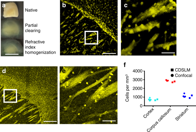
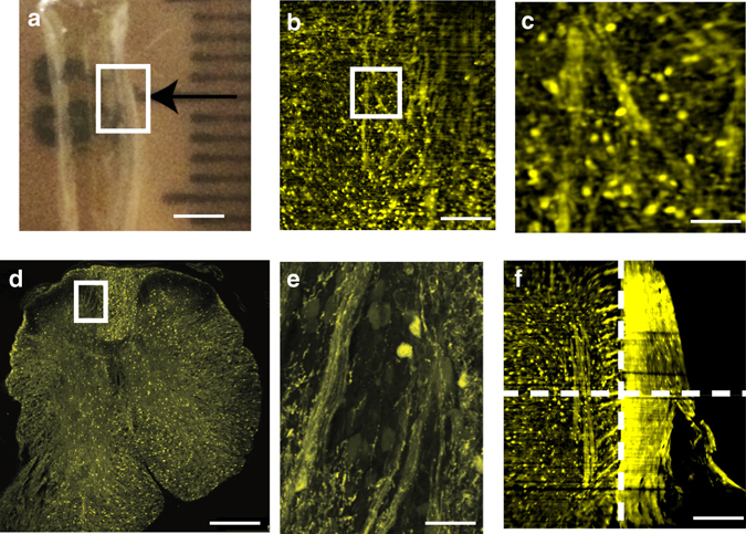
Optical tissue clearing has revolutionized researchers' ability to perform fluorescent measurements of molecules, cells, and structures within intact tissue. One common complication to all optically cleared tissue is a spatially heterogeneous refractive index, leading to light scattering and first-order defocus. We designed C-DSLM (cleared tissue digital scanned light-sheet microscopy) as a low-cost method intended to automatically generate in-focus images of cleared tissue. We demonstrate the flexibility and power of C-DSLM by quantifying fluorescent features in tissue from multiple animal models using refractive index matched and mismatched microscope objectives. This includes a unique measurement of myelin tracks within intact tissue using an endogenous fluorescent reporter where typical clearing approaches render such structures difficult to image. For all measurements, we provide independent verification using standard serial tissue sectioning and quantification methods. Paired with advancements in volumetric image processing, C-DSLM provides a robust methodology to quantify sub-micron features within large tissue sections.Optical clearing of tissue has enabled optical imaging deeper into tissue due to significantly reduced light scattering. Here, Ryan et al. tackle first-order defocus, an artefact of a non-uniform refractive index, extending light-sheet microscopy to partially cleared samples.

摘要

光学组织透明化技术彻底改变了研究人员对完整组织内分子、细胞和结构进行荧光测量的能力。所有光学透明化组织都存在一个常见的并发症,即空间异质的折射率,这会导致光散射和一阶散焦。我们设计了C-DSLM(透明组织数字扫描光片显微镜)作为一种低成本方法,旨在自动生成透明组织的聚焦图像。我们通过使用折射率匹配和不匹配的显微镜物镜对多个动物模型的组织中的荧光特征进行量化,展示了C-DSLM的灵活性和强大功能。这包括使用内源性荧光报告分子对完整组织内的髓磷脂轨迹进行独特测量,而典型的透明化方法使此类结构难以成像。对于所有测量,我们使用标准的连续组织切片和量化方法进行独立验证。与体积图像处理的进展相结合,C-DSLM提供了一种强大的方法来量化大组织切片内的亚微米特征。由于光散射显著减少,组织的光学透明化使得光学成像能够更深入组织。在此,瑞安等人解决了一阶散焦问题,这是一种非均匀折射率的假象,将光片显微镜扩展到部分透明的样本。

https://cdn.ncbi.nlm.nih.gov/pmc/blobs/7f72/5606987/a918e0aa456d/41467_2017_514_Fig6_HTML.jpg
https://cdn.ncbi.nlm.nih.gov/pmc/blobs/7f72/5606987/1f6201b6743b/41467_2017_514_Fig1_HTML.jpg
https://cdn.ncbi.nlm.nih.gov/pmc/blobs/7f72/5606987/3df15544c252/41467_2017_514_Fig2_HTML.jpg
https://cdn.ncbi.nlm.nih.gov/pmc/blobs/7f72/5606987/f3b31b7007c3/41467_2017_514_Fig3_HTML.jpg
https://cdn.ncbi.nlm.nih.gov/pmc/blobs/7f72/5606987/00ef85c1dcfc/41467_2017_514_Fig4_HTML.jpg
https://cdn.ncbi.nlm.nih.gov/pmc/blobs/7f72/5606987/86789712fb31/41467_2017_514_Fig5_HTML.jpg
https://cdn.ncbi.nlm.nih.gov/pmc/blobs/7f72/5606987/a918e0aa456d/41467_2017_514_Fig6_HTML.jpg

相似文献

1
Automatic and adaptive heterogeneous refractive index compensation for light-sheet microscopy.用于光片显微镜的自动自适应异质折射率补偿
Nat Commun. 2017 Sep 20;8(1):612. doi: 10.1038/s41467-017-00514-7.
2
High-resolution in-depth imaging of optically cleared thick samples using an adaptive SPIM.使用自适应光片显微镜对光学透明厚样品进行高分辨率深度成像。
Sci Rep. 2015 Nov 18;5:16898. doi: 10.1038/srep16898.
3
3D Imaging for Cleared Tissues and Thicker Samples on Confocal and Light-Sheet Microscopes.共聚焦显微镜和光片显微镜对透明组织和较厚样本的三维成像
Methods Mol Biol. 2023;2593:143-161. doi: 10.1007/978-1-0716-2811-9_9.
4
Single-Step Fast Tissue Clearing of Thick Mouse Brain Tissue for Multi-Dimensional High-Resolution Imaging.一步法快速透明化厚鼠脑组织以进行多维高分辨率成像。
Int J Mol Sci. 2022 Jun 19;23(12):6826. doi: 10.3390/ijms23126826.
5
Scanning light-sheet microscopy in the whole mouse brain with HiLo background rejection.基于 HiLo 背景抑制的全鼠脑扫描光片显微镜成像
J Biomed Opt. 2010 Jan-Feb;15(1):016027. doi: 10.1117/1.3324890.
6
Digital scanned laser light-sheet fluorescence lifetime microscopy with wide-field time-gated imaging.数字扫描激光片层荧光寿命显微镜与宽场时间门成像。
J Microsc. 2020 Jul;279(1):69-76. doi: 10.1111/jmi.12898. Epub 2020 May 14.
7
Three-dimensional refractive index reconstruction with quantitative phase tomography.基于定量相位断层扫描的三维折射率重建
Microsc Res Tech. 2008 Jan;71(1):5-10. doi: 10.1002/jemt.20520.
8
Whole-Brain Single-Cell Imaging and Analysis of Intact Neonatal Mouse Brains Using MRI, Tissue Clearing, and Light-Sheet Microscopy.利用 MRI、组织透明化和光片显微镜对完整新生鼠脑进行全脑单细胞成像和分析。
J Vis Exp. 2022 Aug 1(186). doi: 10.3791/64096.
9
Sample Preparation of Optically Cleared Liver Tissue to Identify Liver Macrophages Using 3D Microscopy.光学透明化肝脏组织样本制备方法,用于使用 3D 显微镜鉴定肝脏中的巨噬细胞。
Methods Mol Biol. 2020;2164:55-63. doi: 10.1007/978-1-0716-0704-6_7.
10
Thin-sheet laser imaging microscopy for optical sectioning of thick tissues.用于厚组织光学切片的薄片激光成像显微镜术。
Biotechniques. 2009 Apr;46(4):287-94. doi: 10.2144/000113087.

引用本文的文献

1
Zebrafish fluorescence imaging platform based on Bessel light sheet illumination.基于贝塞尔光片照明的斑马鱼荧光成像平台。
Biomed Opt Express. 2025 Mar 27;16(4):1678-1691. doi: 10.1364/BOE.542599. eCollection 2025 Apr 1.
2
A multiwell plate approach to increase the sample throughput during tissue clearing.一种用于在组织透明化过程中提高样品通量的多孔板方法。
Nat Protoc. 2025 Apr;20(4):967-988. doi: 10.1038/s41596-024-01080-1. Epub 2024 Dec 3.
3
Full-aperture extended-depth oblique plane microscopy through dynamic remote focusing.

本文引用的文献

1
Quantifying three-dimensional rodent retina vascular development using optical tissue clearing and light-sheet microscopy.利用光学组织透明化和光片显微镜定量研究三维啮齿动物视网膜血管发育。
J Biomed Opt. 2017 Jul 1;22(7):76011. doi: 10.1117/1.JBO.22.7.076011.
2
Selectable light-sheet uniformity using tuned axial scanning.使用调谐轴向扫描实现可选择的光片均匀性。
Microsc Res Tech. 2017 Feb;80(2):250-259. doi: 10.1002/jemt.22795. Epub 2016 Nov 6.
3
A beginner's guide to tissue clearing.组织透明化新手指南。
全孔径扩展景深斜平面显微镜通过动态远程聚焦。
J Biomed Opt. 2024 Mar;29(3):036502. doi: 10.1117/1.JBO.29.3.036502. Epub 2024 Mar 21.
4
Volumetric light sheet imaging with adaptive optics correction.具有自适应光学校正的体积光片成像
Biomed Opt Express. 2023 Mar 29;14(4):1757-1771. doi: 10.1364/BOE.473237. eCollection 2023 Apr 1.
5
Deep learning-based autofocus method enhances image quality in light-sheet fluorescence microscopy.基于深度学习的自动对焦方法提高了光片荧光显微镜的图像质量。
Biomed Opt Express. 2021 Jul 22;12(8):5214-5226. doi: 10.1364/BOE.427099. eCollection 2021 Aug 1.
6
Whole brain functional recordings at cellular resolution in zebrafish larvae with 3D scanning multiphoton microscopy.利用 3D 扫描双光子显微镜在斑马鱼幼虫中进行细胞分辨率的全脑功能记录。
Sci Rep. 2021 May 26;11(1):11048. doi: 10.1038/s41598-021-90335-y.
7
Tutorial: practical considerations for tissue clearing and imaging.教程:组织透明化和成像的实际考量
Nat Protoc. 2021 Jun;16(6):2732-2748. doi: 10.1038/s41596-021-00502-8. Epub 2021 May 21.
8
Tissue clearing and its applications in neuroscience.组织透明化及其在神经科学中的应用。
Nat Rev Neurosci. 2020 Feb;21(2):61-79. doi: 10.1038/s41583-019-0250-1.
9
Hybrid light-sheet and light-field microscope for high resolution and large volume neuroimaging.用于高分辨率和大体积神经成像的混合光片和光场显微镜。
Biomed Opt Express. 2019 Nov 27;10(12):6595-6610. doi: 10.1364/BOE.10.006595. eCollection 2019 Dec 1.
10
Advanced CUBIC tissue clearing for whole-organ cell profiling.高级 CUBIC 组织通透化技术用于整体器官细胞剖析。
Nat Protoc. 2019 Dec;14(12):3506-3537. doi: 10.1038/s41596-019-0240-9. Epub 2019 Nov 20.
Int J Biochem Cell Biol. 2017 Mar;84:35-39. doi: 10.1016/j.biocel.2016.12.009. Epub 2017 Jan 7.
4
Adaptive light-sheet microscopy for long-term, high-resolution imaging in living organisms.自适应光片显微镜用于活体生物的长期、高分辨率成像。
Nat Biotechnol. 2016 Dec;34(12):1267-1278. doi: 10.1038/nbt.3708. Epub 2016 Oct 31.
5
Selective plane illumination microscopy with a light sheet of uniform thickness formed by an electrically tunable lens.利用电可调透镜形成均匀厚度光片的选择性平面照明显微术。
Microsc Res Tech. 2018 Sep;81(9):924-928. doi: 10.1002/jemt.22707. Epub 2016 Jun 24.
6
Chemical Principles in Tissue Clearing and Staining Protocols for Whole-Body Cell Profiling.化学原理在组织通透和染色技术中的应用,用于整体细胞分析。
Annu Rev Cell Dev Biol. 2016 Oct 6;32:713-741. doi: 10.1146/annurev-cellbio-111315-125001. Epub 2016 Jun 8.
7
Diagonally Scanned Light-Sheet Microscopy for Fast Volumetric Imaging of Adherent Cells.用于贴壁细胞快速体积成像的对角扫描光片显微镜术
Biophys J. 2016 Mar 29;110(6):1456-65. doi: 10.1016/j.bpj.2016.01.029.
8
Pushing Forward: Remyelination as the New Frontier in CNS Diseases.推动前进:中枢神经系统疾病中的髓鞘修复新前沿。
Trends Neurosci. 2016 Apr;39(4):246-263. doi: 10.1016/j.tins.2016.02.004. Epub 2016 Mar 7.
9
TeraFly: real-time three-dimensional visualization and annotation of terabytes of multidimensional volumetric images.TeraFly:数万亿字节多维体图像的实时三维可视化与标注
Nat Methods. 2016 Mar;13(3):192-4. doi: 10.1038/nmeth.3767.
10
Remodeling myelination: implications for mechanisms of neural plasticity.重塑髓鞘形成:对神经可塑性机制的影响
Nat Neurosci. 2016 Feb;19(2):190-7. doi: 10.1038/nn.4200.