Beck S, Gunawardena P, Horton D L, Hicks D J, Marston D A, Ortiz-Pelaez A, Fooks A R, Núñez A
Pathology Department, Animal and Plant Health Agency, Weybridge, UK.
Department of Veterinary Pathobiology, University of Peradeniya, Peradeniya, Sri Lanka.
BMC Vet Res. 2017 Apr 12;13(1):99. doi: 10.1186/s12917-017-1024-5.
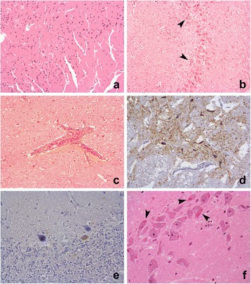
The recommended screening of rabies in 'suspect' animal cases involves testing fresh brain tissue. The preservation of fresh tissue however can be difficult under field conditions and formalin fixation provides a simple alternative that may allow a confirmatory diagnosis. The occurrence and location of histopathological changes and immunohistochemical (IHC) labelling for rabies in formalin fixed paraffin embedded (FFPE) canine brain is described in samples from 57 rabies suspect cases from Sri-Lanka. The presence of Negri bodies and immunohistochemical detection of rabies virus antigen were evaluated in the cortex, hippocampus, cerebellum and brainstem. The effect of autolysis and artefactual degeneration of the tissue was also assessed.
Rabies was confirmed in 53 of 57 (93%) cases by IHC. IHC labelling was statistically more abundant in the brainstem. Negri bodies were observed in 32 of 53 (60.4%) of the positive cases. Although tissue degradation had no effect on IHC diagnosis, it was associated with an inability to detect Negri bodies. In 13 cases, a confirmatory Polymerase chain reaction (PCR) testing for rabies virus RNA was undertaken by extracting RNA from fresh frozen tissue, and also attempted using FFPE samples. PCR detection using fresh frozen samples was in agreement with the IHC results. The PCR method from FFPE tissues was suitable for control material but unsuccessful in our field cases.
Histopathological examination of the brain is essential to define the differential diagnoses of behaviour modifying conditions in rabies virus negative cases, but it is unreliable as the sole method for rabies diagnosis, particularly where artefactual change has occurred. Formalin fixation and paraffin embedding does not prevent detection of rabies virus via IHC labelling even where artefactual degeneration has occurred. This could represent a pragmatic secondary assay for rabies diagnosis in the field because formalin fixation can prevent sample degeneration. The brain stem was shown to be the site with most viral immunoreactivity; supporting recommended sampling protocols in favour of improved necropsy safety in the field. PCR testing of formalin fixed tissue may be successful in certain circumstances as an alternative test.
在“疑似”动物病例中,狂犬病的推荐筛查方法是检测新鲜脑组织。然而,在野外条件下保存新鲜组织可能很困难,而福尔马林固定提供了一种简单的替代方法,可能有助于确诊。本文描述了来自斯里兰卡的57例狂犬病疑似病例样本中,福尔马林固定石蜡包埋(FFPE)犬脑组织中狂犬病组织病理学变化和免疫组织化学(IHC)标记的发生情况及位置。在皮质、海马体、小脑和脑干中评估了内基小体的存在以及狂犬病病毒抗原的免疫组织化学检测情况。还评估了组织自溶和人为变性的影响。
通过免疫组织化学方法,在57例病例中的53例(93%)确诊为狂犬病。免疫组织化学标记在脑干中的数量在统计学上更多。在53例阳性病例中的32例(60.4%)观察到了内基小体。尽管组织降解对免疫组织化学诊断没有影响,但它与无法检测到内基小体有关。在13例病例中,通过从新鲜冷冻组织中提取RNA进行了狂犬病病毒RNA的验证性聚合酶链反应(PCR)检测,也尝试使用FFPE样本进行检测。使用新鲜冷冻样本的PCR检测结果与免疫组织化学结果一致。来自FFPE组织的PCR方法适用于对照材料,但在我们的野外病例中未成功。
对大脑进行组织病理学检查对于明确狂犬病病毒阴性病例中行为改变状况的鉴别诊断至关重要,但作为狂犬病诊断的唯一方法并不可靠,特别是在发生人为变化的情况下。即使发生了人为变性,福尔马林固定和石蜡包埋也不会妨碍通过免疫组织化学标记检测狂犬病病毒。这可能代表了一种在野外用于狂犬病诊断的实用二级检测方法,因为福尔马林固定可以防止样本变性。脑干被证明是病毒免疫反应性最强的部位;支持推荐的采样方案,以提高野外尸检的安全性。在某些情况下,对福尔马林固定组织进行PCR检测作为替代检测方法可能会成功。