• 文献检索
  • 文档翻译
  • 深度研究
  • 学术资讯
  • Suppr Zotero 插件Zotero 插件
  • 邀请有礼
  • 套餐&价格
  • 历史记录
应用&插件
Suppr Zotero 插件Zotero 插件浏览器插件Mac 客户端Windows 客户端微信小程序
定价
高级版会员购买积分包购买API积分包
服务
文献检索文档翻译深度研究API 文档MCP 服务
关于我们
关于 Suppr公司介绍联系我们用户协议隐私条款
关注我们

Suppr 超能文献

核心技术专利:CN118964589B侵权必究
粤ICP备2023148730 号-1Suppr @ 2026

文献检索

告别复杂PubMed语法,用中文像聊天一样搜索,搜遍4000万医学文献。AI智能推荐,让科研检索更轻松。

立即免费搜索

文件翻译

保留排版,准确专业,支持PDF/Word/PPT等文件格式,支持 12+语言互译。

免费翻译文档

深度研究

AI帮你快速写综述,25分钟生成高质量综述,智能提取关键信息,辅助科研写作。

立即免费体验

基于概率荧光的突触检测。

Probabilistic fluorescence-based synapse detection.

作者信息

Simhal Anish K, Aguerrebere Cecilia, Collman Forrest, Vogelstein Joshua T, Micheva Kristina D, Weinberg Richard J, Smith Stephen J, Sapiro Guillermo

机构信息

Electrical and Computer Engineering, Duke University, Durham, North Carolina, United States of America.

Synapse Biology, Allen Institute for Brain Sciences, Seattle, Washington, United States of America.

出版信息

PLoS Comput Biol. 2017 Apr 17;13(4):e1005493. doi: 10.1371/journal.pcbi.1005493. eCollection 2017 Apr.

DOI:10.1371/journal.pcbi.1005493
PMID:28414801
原文链接:https://pmc.ncbi.nlm.nih.gov/articles/PMC5411093/
Abstract

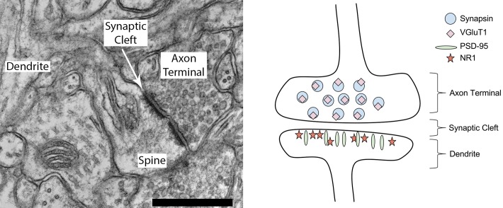
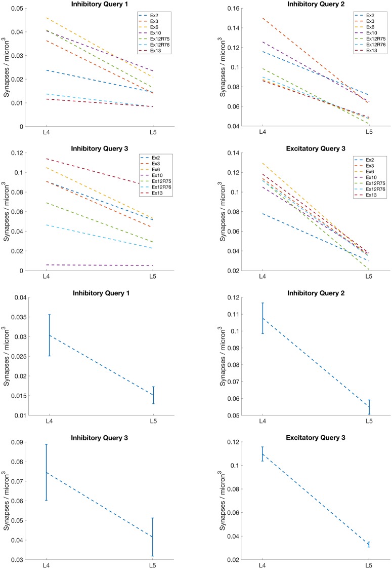
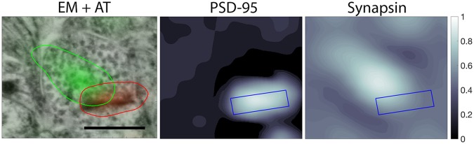
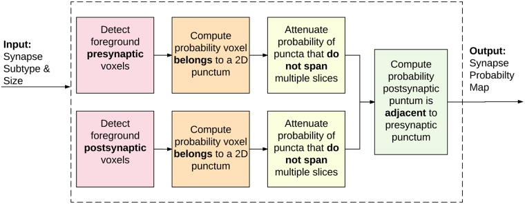
Deeper exploration of the brain's vast synaptic networks will require new tools for high-throughput structural and molecular profiling of the diverse populations of synapses that compose those networks. Fluorescence microscopy (FM) and electron microscopy (EM) offer complementary advantages and disadvantages for single-synapse analysis. FM combines exquisite molecular discrimination capacities with high speed and low cost, but rigorous discrimination between synaptic and non-synaptic fluorescence signals is challenging. In contrast, EM remains the gold standard for reliable identification of a synapse, but offers only limited molecular discrimination and is slow and costly. To develop and test single-synapse image analysis methods, we have used datasets from conjugate array tomography (cAT), which provides voxel-conjugate FM and EM (annotated) images of the same individual synapses. We report a novel unsupervised probabilistic method for detection of synapses from multiplex FM (muxFM) image data, and evaluate this method both by comparison to EM gold standard annotated data and by examining its capacity to reproduce known important features of cortical synapse distributions. The proposed probabilistic model-based synapse detector accepts molecular-morphological synapse models as user queries, and delivers a volumetric map of the probability that each voxel represents part of a synapse. Taking human annotation of cAT EM data as ground truth, we show that our algorithm detects synapses from muxFM data alone as successfully as human annotators seeing only the muxFM data, and accurately reproduces known architectural features of cortical synapse distributions. This approach opens the door to data-driven discovery of new synapse types and their density. We suggest that our probabilistic synapse detector will also be useful for analysis of standard confocal and super-resolution FM images, where EM cross-validation is not practical.

摘要

要更深入地探索大脑庞大的突触网络,将需要新的工具,以便对构成这些网络的各种突触群体进行高通量的结构和分子分析。荧光显微镜(FM)和电子显微镜(EM)在单突触分析方面各有优缺点。FM结合了出色的分子辨别能力、高速度和低成本,但在突触荧光信号和非突触荧光信号之间进行严格区分具有挑战性。相比之下,EM仍然是可靠识别突触的金标准,但分子辨别能力有限,且速度慢、成本高。为了开发和测试单突触图像分析方法,我们使用了共轭阵列断层扫描(cAT)的数据集,该数据集提供了同一单个突触的体素共轭FM和EM(注释)图像。我们报告了一种从多通道FM(muxFM)图像数据中检测突触的新型无监督概率方法,并通过与EM金标准注释数据进行比较以及检查其再现皮质突触分布已知重要特征的能力来评估该方法。所提出的基于概率模型的突触检测器接受分子形态突触模型作为用户查询,并给出每个体素代表突触一部分的概率的体积图。以cAT EM数据的人工注释作为基准事实,我们表明我们的算法仅从muxFM数据中检测突触的成功率与仅查看muxFM数据的人工注释者相同,并且准确地再现了皮质突触分布的已知结构特征。这种方法为数据驱动的新突触类型及其密度的发现打开了大门。我们认为,我们的概率突触检测器对于标准共聚焦和超分辨率FM图像的分析也将是有用的,在这些图像中EM交叉验证不实用。

https://cdn.ncbi.nlm.nih.gov/pmc/blobs/328f/5411093/3bbf920b6a0d/pcbi.1005493.g016.jpg
https://cdn.ncbi.nlm.nih.gov/pmc/blobs/328f/5411093/899a55585866/pcbi.1005493.g001.jpg
https://cdn.ncbi.nlm.nih.gov/pmc/blobs/328f/5411093/4c435ee135fa/pcbi.1005493.g002.jpg
https://cdn.ncbi.nlm.nih.gov/pmc/blobs/328f/5411093/94d215a43a45/pcbi.1005493.g003.jpg
https://cdn.ncbi.nlm.nih.gov/pmc/blobs/328f/5411093/d4da506a1892/pcbi.1005493.g004.jpg
https://cdn.ncbi.nlm.nih.gov/pmc/blobs/328f/5411093/ab4fbeae3e4f/pcbi.1005493.g005.jpg
https://cdn.ncbi.nlm.nih.gov/pmc/blobs/328f/5411093/3d19fb1c24ad/pcbi.1005493.g006.jpg
https://cdn.ncbi.nlm.nih.gov/pmc/blobs/328f/5411093/693df64c90cb/pcbi.1005493.g007.jpg
https://cdn.ncbi.nlm.nih.gov/pmc/blobs/328f/5411093/3e260332a2a0/pcbi.1005493.g008.jpg
https://cdn.ncbi.nlm.nih.gov/pmc/blobs/328f/5411093/465b120eb91b/pcbi.1005493.g009.jpg
https://cdn.ncbi.nlm.nih.gov/pmc/blobs/328f/5411093/06a9f537269e/pcbi.1005493.g010.jpg
https://cdn.ncbi.nlm.nih.gov/pmc/blobs/328f/5411093/199a15a5091e/pcbi.1005493.g011.jpg
https://cdn.ncbi.nlm.nih.gov/pmc/blobs/328f/5411093/37772de951a9/pcbi.1005493.g012.jpg
https://cdn.ncbi.nlm.nih.gov/pmc/blobs/328f/5411093/11842def7b5d/pcbi.1005493.g013.jpg
https://cdn.ncbi.nlm.nih.gov/pmc/blobs/328f/5411093/6ed81d72a562/pcbi.1005493.g014.jpg
https://cdn.ncbi.nlm.nih.gov/pmc/blobs/328f/5411093/7b7966cfa74f/pcbi.1005493.g015.jpg
https://cdn.ncbi.nlm.nih.gov/pmc/blobs/328f/5411093/3bbf920b6a0d/pcbi.1005493.g016.jpg

相似文献

1
Probabilistic fluorescence-based synapse detection.基于概率荧光的突触检测。
PLoS Comput Biol. 2017 Apr 17;13(4):e1005493. doi: 10.1371/journal.pcbi.1005493. eCollection 2017 Apr.
2
Mapping synapses by conjugate light-electron array tomography.通过共轭光电子阵列断层扫描绘制突触图谱。
J Neurosci. 2015 Apr 8;35(14):5792-807. doi: 10.1523/JNEUROSCI.4274-14.2015.
3
Learning context cues for synapse segmentation.学习突触分割的上下文线索。
IEEE Trans Med Imaging. 2013 Oct;32(10):1864-77. doi: 10.1109/TMI.2013.2267747. Epub 2013 Jun 11.
4
DoGNet: A deep architecture for synapse detection in multiplexed fluorescence images.DoGNet:一种用于多路复用荧光图像中突触检测的深度架构。
PLoS Comput Biol. 2019 May 13;15(5):e1007012. doi: 10.1371/journal.pcbi.1007012. eCollection 2019 May.
5
Sub-synaptic, multiplexed analysis of proteins reveals Fragile X related protein 2 is mislocalized in KO synapses.蛋白质的突触下多重分析表明脆性X相关蛋白2在基因敲除突触中定位错误。
Elife. 2016 Oct 22;5:e20560. doi: 10.7554/eLife.20560.
6
Automated analysis of a diverse synapse population.多样化突触群体的自动化分析。
PLoS Comput Biol. 2013;9(3):e1002976. doi: 10.1371/journal.pcbi.1002976. Epub 2013 Mar 28.
7
Automated detection of synapses in serial section transmission electron microscopy image stacks.在连续切片透射电子显微镜图像堆栈中自动检测突触。
PLoS One. 2014 Feb 6;9(2):e87351. doi: 10.1371/journal.pone.0087351. eCollection 2014.
8
RoboEM: automated 3D flight tracing for synaptic-resolution connectomics.RoboEM:用于突触分辨率连接组学的自动化 3D 飞行追踪
Nat Methods. 2024 May;21(5):908-913. doi: 10.1038/s41592-024-02226-5. Epub 2024 Mar 21.
9
Fully-Automatic Synapse Prediction and Validation on a Large Data Set.全自动突触预测及其在大数据集上的验证。
Front Neural Circuits. 2018 Oct 29;12:87. doi: 10.3389/fncir.2018.00087. eCollection 2018.
10
Learning context cues for synapse segmentation in EM volumes.在电子显微镜图像中学习用于突触分割的上下文线索。
Med Image Comput Comput Assist Interv. 2012;15(Pt 1):585-92. doi: 10.1007/978-3-642-33415-3_72.

引用本文的文献

1
Data-driven synapse classification reveals a logic of glutamate receptor diversity.数据驱动的突触分类揭示了谷氨酸受体多样性的逻辑。
bioRxiv. 2025 Jan 14:2024.12.11.628056. doi: 10.1101/2024.12.11.628056.
2
Developing a Toolbox of Antibodies Validated for Array Tomography-Based Imaging of Brain Synapses.开发一套经阵列层析成像技术验证的用于检测脑突触的抗体工具。
eNeuro. 2023 Dec 22;10(12). doi: 10.1523/ENEURO.0290-23.2023. Print 2023 Dec.
3
Developing a Toolbox of Antibodies Validated for Array Tomography-Based Imaging of Brain Synapses.

本文引用的文献

1
Noise-induced cochlear synaptopathy: Past findings and future studies.噪声性耳蜗突触病变:既往研究结果与未来研究方向
Hear Res. 2017 Jun;349:148-154. doi: 10.1016/j.heares.2016.12.008. Epub 2016 Dec 19.
2
High-performance multiplexed fluorescence in situ hybridization in culture and tissue with matrix imprinting and clearing.利用基质印记和透明化技术在培养物和组织中进行的高性能多重荧光原位杂交。
Proc Natl Acad Sci U S A. 2016 Dec 13;113(50):14456-14461. doi: 10.1073/pnas.1617699113. Epub 2016 Nov 22.
3
Sub-synaptic, multiplexed analysis of proteins reveals Fragile X related protein 2 is mislocalized in KO synapses.
开发用于基于阵列断层扫描的脑突触成像的经过验证的抗体工具箱。
bioRxiv. 2023 Nov 8:2023.06.28.546920. doi: 10.1101/2023.06.28.546920.
4
GABA bouton subpopulations in the human dentate gyrus are differentially altered in mesial temporal lobe epilepsy.人类齿状回中的 GABA 末梢亚群在颞叶内侧癫痫中存在差异改变。
J Neurophysiol. 2020 Jan 1;123(1):392-406. doi: 10.1152/jn.00523.2018. Epub 2019 Dec 4.
5
SynQuant: an automatic tool to quantify synapses from microscopy images.SynQuant:一种自动工具,用于从显微镜图像中定量突触。
Bioinformatics. 2020 Mar 1;36(5):1599-1606. doi: 10.1093/bioinformatics/btz760.
6
Multifaceted Changes in Synaptic Composition and Astrocytic Involvement in a Mouse Model of Fragile X Syndrome.脆性 X 综合征小鼠模型中突触组成的多方面变化和星形胶质细胞的参与。
Sci Rep. 2019 Sep 25;9(1):13855. doi: 10.1038/s41598-019-50240-x.
7
DoGNet: A deep architecture for synapse detection in multiplexed fluorescence images.DoGNet:一种用于多路复用荧光图像中突触检测的深度架构。
PLoS Comput Biol. 2019 May 13;15(5):e1007012. doi: 10.1371/journal.pcbi.1007012. eCollection 2019 May.
8
A community-developed open-source computational ecosystem for big neuro data.一个由社区开发的用于处理大规模神经数据的开源计算生态系统。
Nat Methods. 2018 Nov;15(11):846-847. doi: 10.1038/s41592-018-0181-1.
9
Q&A: Array tomography.常见问题解答:面绘制技术
BMC Biol. 2018 Sep 6;16(1):98. doi: 10.1186/s12915-018-0560-1.
10
Development of a Novel FIJI-Based Method to Investigate Neuronal Circuitry in Neonatal Mice.开发一种基于 FIJI 的新方法来研究新生小鼠的神经元回路。
Dev Neurobiol. 2018 Nov;78(11):1146-1167. doi: 10.1002/dneu.22636. Epub 2018 Sep 20.
蛋白质的突触下多重分析表明脆性X相关蛋白2在基因敲除突触中定位错误。
Elife. 2016 Oct 22;5:e20560. doi: 10.7554/eLife.20560.
4
Mechanisms and functional roles of glutamatergic synapse diversity in a cerebellar circuit.小脑回路中谷氨酸能突触多样性的机制及功能作用
Elife. 2016 Sep 19;5:e15872. doi: 10.7554/eLife.15872.
5
From Cajal to Connectome and Beyond.从 Cajal 到连接组学及超越
Annu Rev Neurosci. 2016 Jul 8;39:197-216. doi: 10.1146/annurev-neuro-071714-033954.
6
Nanoscale imaging of RNA with expansion microscopy.利用扩张显微镜对RNA进行纳米级成像。
Nat Methods. 2016 Aug;13(8):679-84. doi: 10.1038/nmeth.3899. Epub 2016 Jul 4.
7
PSD95 nanoclusters are postsynaptic building blocks in hippocampus circuits.突触后密度蛋白95(PSD95)纳米簇是海马体回路中的突触后构建模块。
Sci Rep. 2016 Apr 25;6:24626. doi: 10.1038/srep24626.
8
Synaptic pathology: A shared mechanism in neurological disease.突触病变:神经疾病的共同机制。
Ageing Res Rev. 2016 Jul;28:72-84. doi: 10.1016/j.arr.2016.04.005. Epub 2016 Apr 20.
9
Structured Dendritic Inhibition Supports Branch-Selective Integration in CA1 Pyramidal Cells.结构型树突抑制支持 CA1 锥体神经元的分支选择性整合。
Neuron. 2016 Mar 2;89(5):1016-30. doi: 10.1016/j.neuron.2016.01.029. Epub 2016 Feb 18.
10
A Fast Method for the Segmentation of Synaptic Junctions and Mitochondria in Serial Electron Microscopic Images of the Brain.一种快速分割脑内连续电子显微镜图像中突触结和线粒体的方法。
Neuroinformatics. 2016 Apr;14(2):235-50. doi: 10.1007/s12021-015-9288-z.