Department of Cellular and Molecular Physiology, Stanford School of Medicine, Stanford, 94305, CA.
Department of Physiology and Membrane Biology, University of California Davis School of Medicine, Davis, 95618, CA.
eNeuro. 2023 Dec 22;10(12). doi: 10.1523/ENEURO.0290-23.2023. Print 2023 Dec.
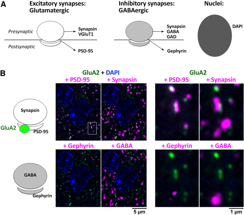
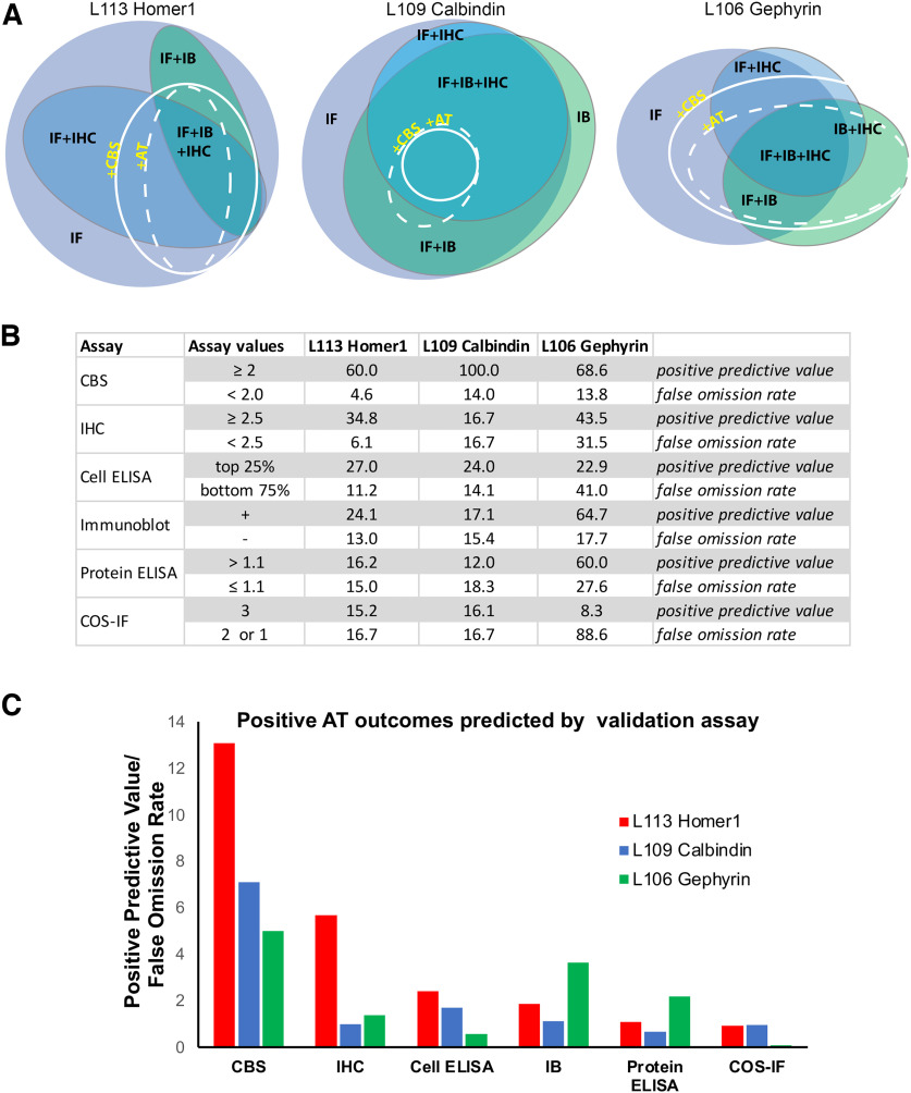
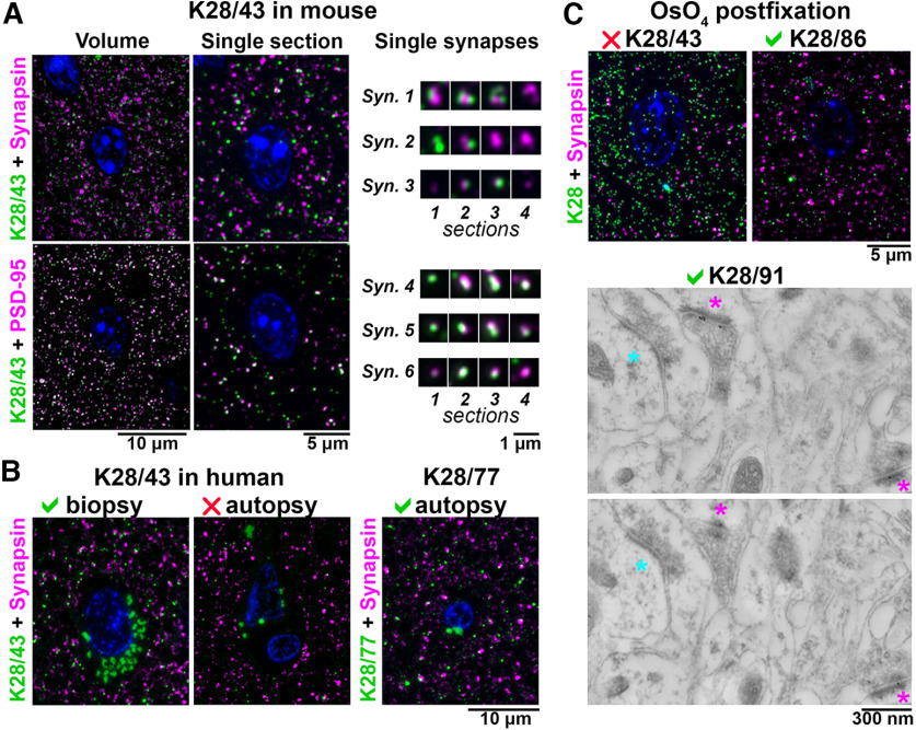
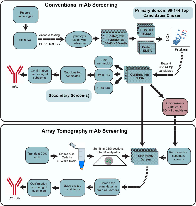
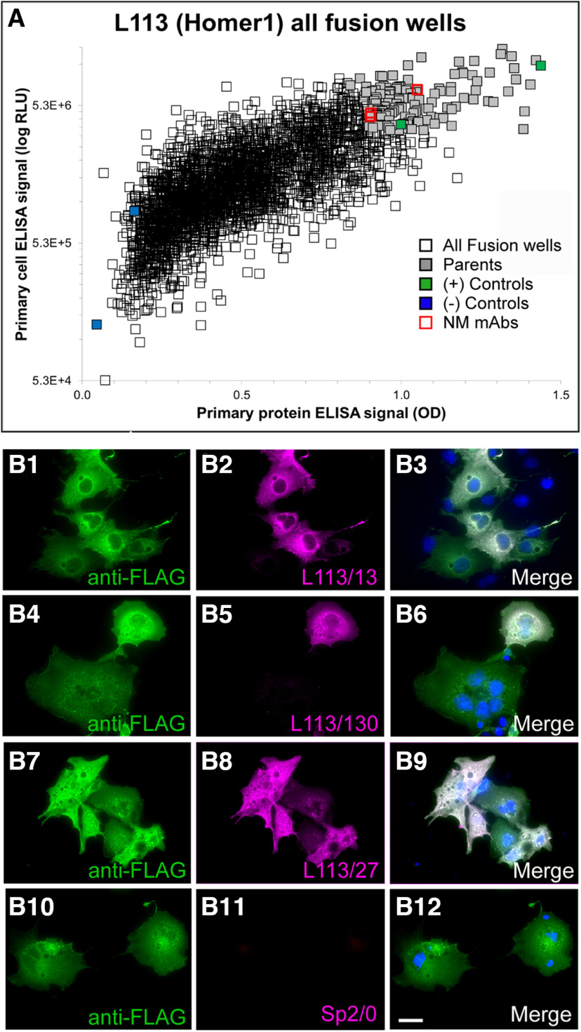
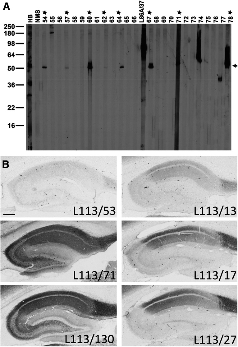
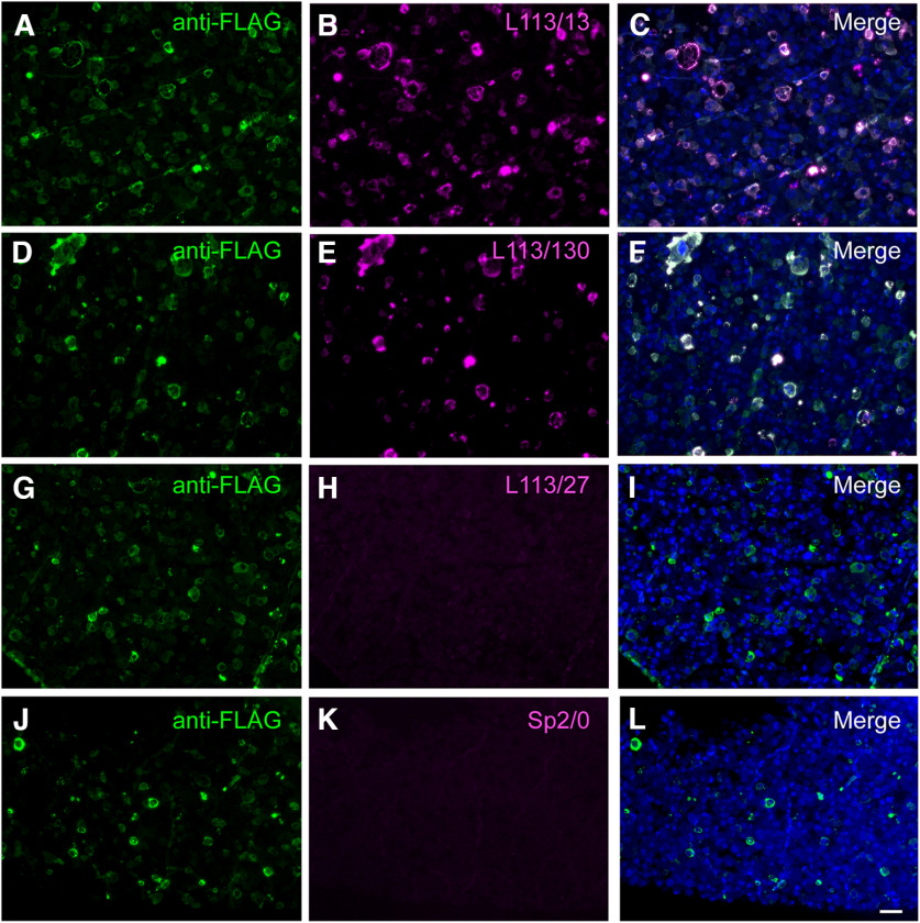
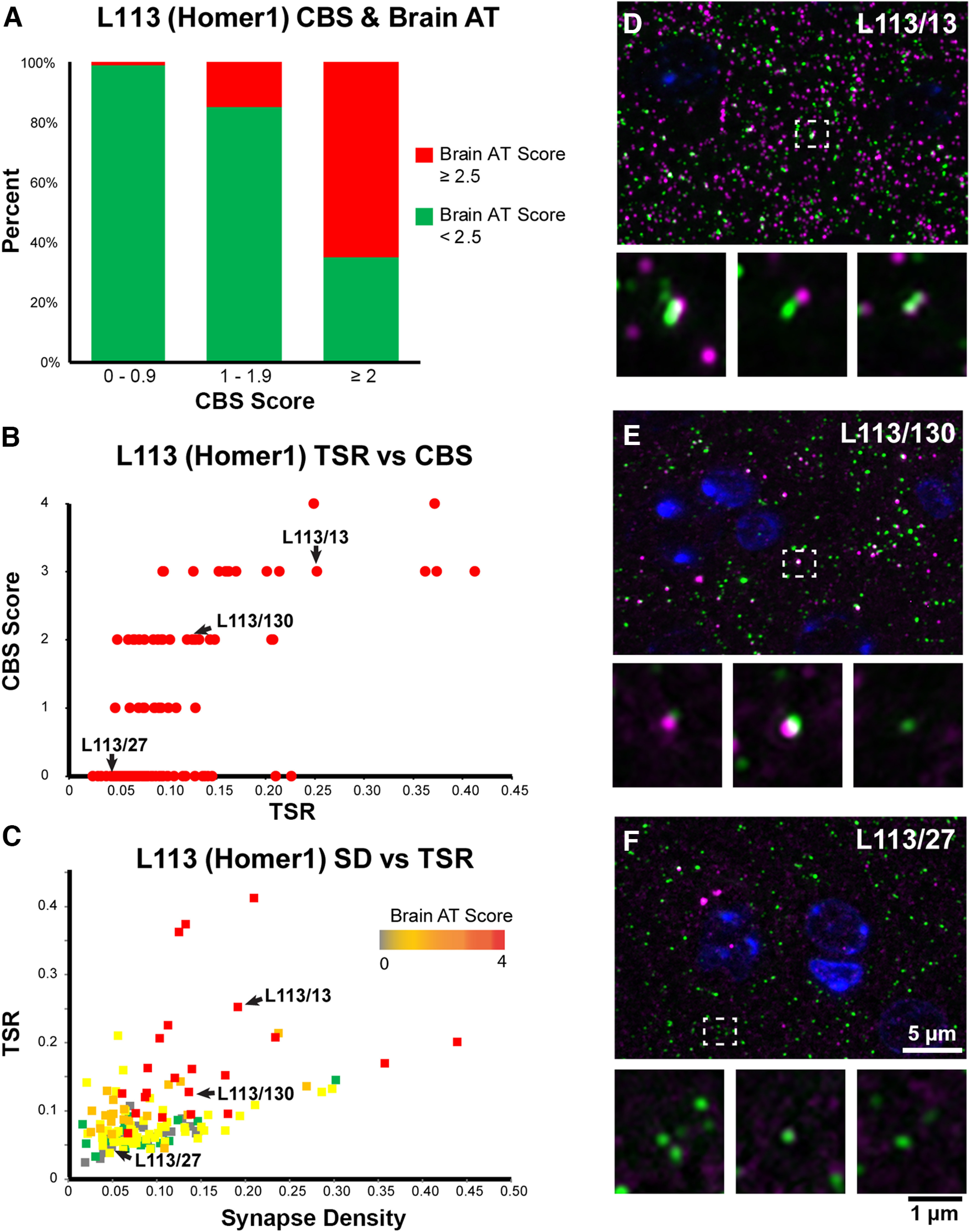
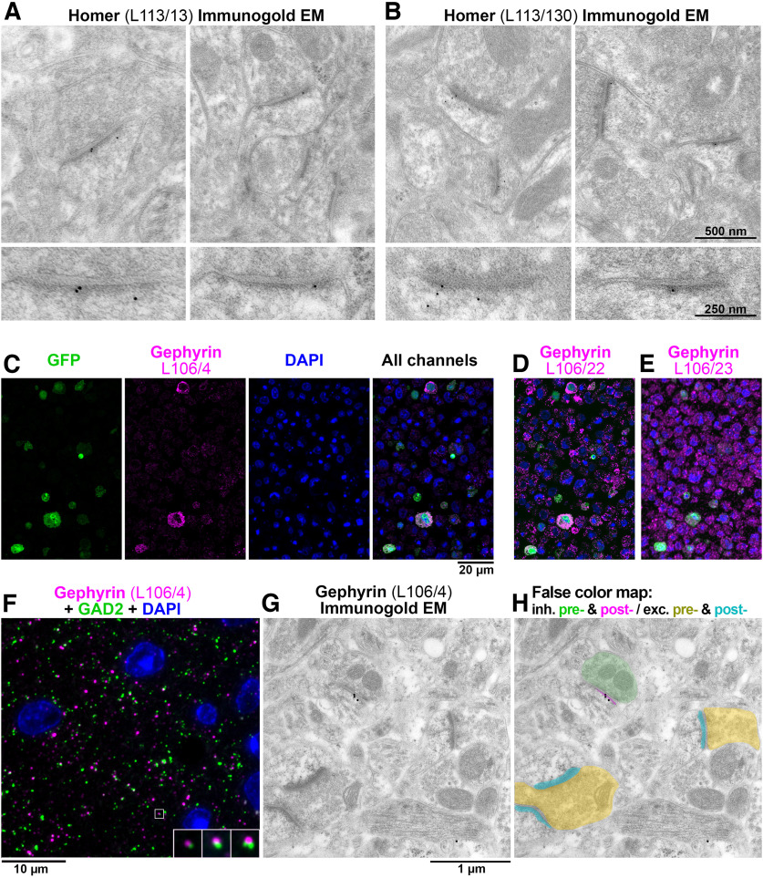
Antibody (Ab)-based imaging techniques rely on reagents whose performance may be application specific. Because commercial antibodies are validated for only a few purposes, users interested in other applications may have to perform extensive in-house antibody testing. Here, we present a novel application-specific proxy screening step to efficiently identify candidate antibodies for array tomography (AT), a serial section volume microscopy technique for high-dimensional quantitative analysis of the cellular proteome. To identify antibodies suitable for AT-based analysis of synapses in mammalian brain, we introduce a heterologous cell-based assay that simulates characteristic features of AT, such as chemical fixation and resin embedding that are likely to influence antibody binding. The assay was included into an initial screening strategy to generate monoclonal antibodies that can be used for AT. This approach simplifies the screening of candidate antibodies and has high predictive value for identifying antibodies suitable for AT analyses. In addition, we have created a comprehensive database of AT-validated antibodies with a neuroscience focus and show that these antibodies have a high likelihood of success for postembedding applications in general, including immunogold electron microscopy. The generation of a large and growing toolbox of AT-compatible antibodies will further enhance the value of this imaging technique.
基于抗体的成像技术依赖于其性能可能因应用而异的试剂。由于商业抗体仅经过少数几种用途的验证,因此对其他应用感兴趣的用户可能不得不进行广泛的内部抗体测试。在这里,我们提出了一种新的特定于应用的代理筛选步骤,以有效地识别用于阵列断层扫描(AT)的候选抗体,AT 是一种用于哺乳动物大脑中突触的高维定量分析的连续切片体积显微镜技术。为了鉴定适用于基于 AT 的哺乳动物大脑中突触分析的抗体,我们引入了一种异源细胞测定法,该测定法模拟了 AT 的特征,例如化学固定和树脂嵌入,这些特征可能会影响抗体结合。该测定法被纳入初始筛选策略中,以生成可用于 AT 的单克隆抗体。这种方法简化了候选抗体的筛选,并且对鉴定适用于 AT 分析的抗体具有很高的预测价值。此外,我们创建了一个具有神经科学重点的针对 AT 验证的抗体的综合数据库,并表明这些抗体在一般的包埋后应用中(包括免疫金电子显微镜)具有很高的成功可能性。生成大量且不断增长的 AT 兼容抗体工具包将进一步提高这种成像技术的价值。