• 文献检索
  • 文档翻译
  • 深度研究
  • 学术资讯
  • Suppr Zotero 插件Zotero 插件
  • 邀请有礼
  • 套餐&价格
  • 历史记录
应用&插件
Suppr Zotero 插件Zotero 插件浏览器插件Mac 客户端Windows 客户端微信小程序
定价
高级版会员购买积分包购买API积分包
服务
文献检索文档翻译深度研究API 文档MCP 服务
关于我们
关于 Suppr公司介绍联系我们用户协议隐私条款
关注我们

Suppr 超能文献

核心技术专利:CN118964589B侵权必究
粤ICP备2023148730 号-1Suppr @ 2026

文献检索

告别复杂PubMed语法,用中文像聊天一样搜索,搜遍4000万医学文献。AI智能推荐,让科研检索更轻松。

立即免费搜索

文件翻译

保留排版,准确专业,支持PDF/Word/PPT等文件格式,支持 12+语言互译。

免费翻译文档

深度研究

AI帮你快速写综述,25分钟生成高质量综述,智能提取关键信息,辅助科研写作。

立即免费体验

超声图像中胰腺癌肝转移的交互式勾画。

Interactive Outlining of Pancreatic Cancer Liver Metastases in Ultrasound Images.

机构信息

Institute for Computer Graphics and Vision, Graz University of Technology, Inffeldgasse 16, 8010, Graz, Austria.

BioTechMed, Krenngasse 37/1, 8010, Graz, Austria.

出版信息

Sci Rep. 2017 Apr 18;7(1):892. doi: 10.1038/s41598-017-00940-z.

DOI:10.1038/s41598-017-00940-z
PMID:28420871
原文链接:https://pmc.ncbi.nlm.nih.gov/articles/PMC5429849/
Abstract

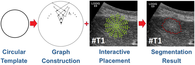
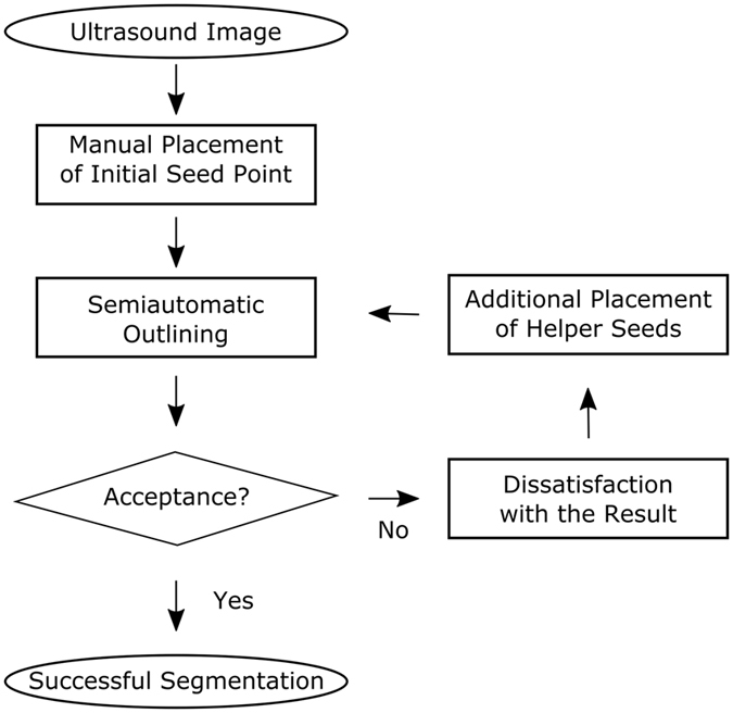
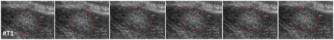
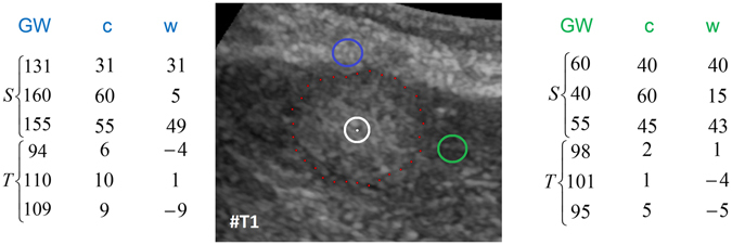
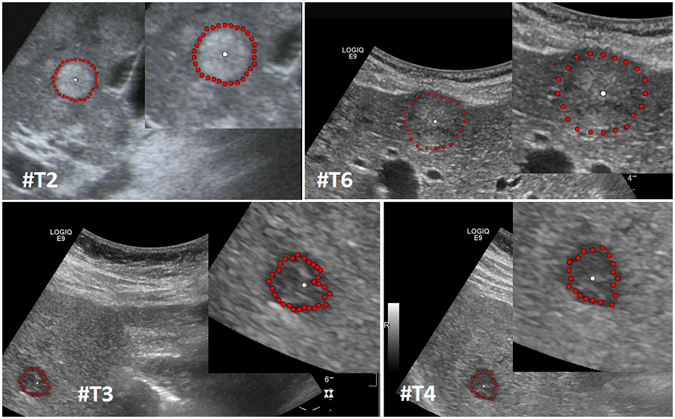
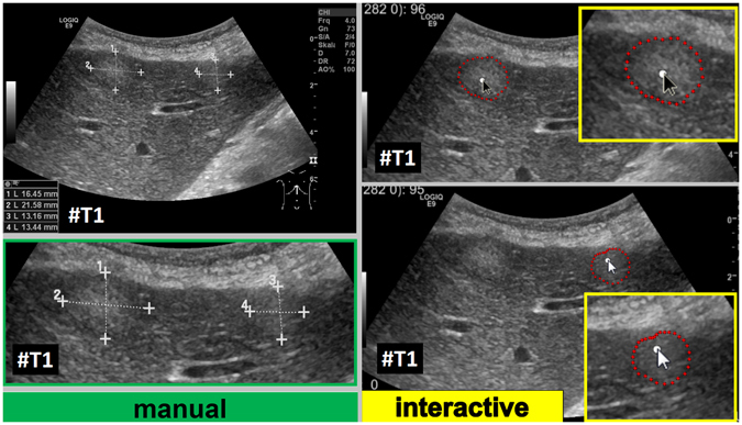
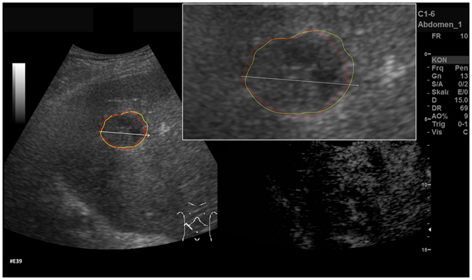
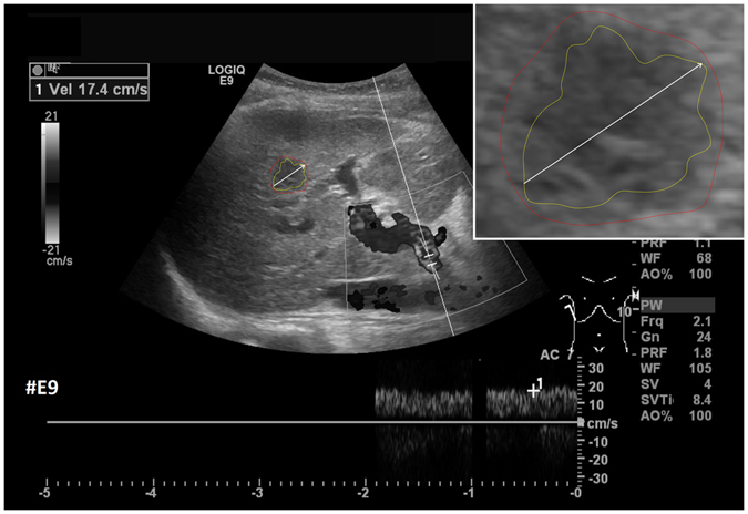
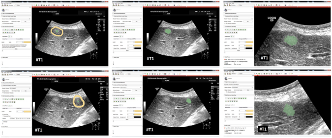
Ultrasound (US) is the most commonly used liver imaging modality worldwide. Due to its low cost, it is increasingly used in the follow-up of cancer patients with metastases localized in the liver. In this contribution, we present the results of an interactive segmentation approach for liver metastases in US acquisitions. A (semi-) automatic segmentation is still very challenging because of the low image quality and the low contrast between the metastasis and the surrounding liver tissue. Thus, the state of the art in clinical practice is still manual measurement and outlining of the metastases in the US images. We tackle the problem by providing an interactive segmentation approach providing real-time feedback of the segmentation results. The approach has been evaluated with typical US acquisitions from the clinical routine, and the datasets consisted of pancreatic cancer metastases. Even for difficult cases, satisfying segmentations results could be achieved because of the interactive real-time behavior of the approach. In total, 40 clinical images have been evaluated with our method by comparing the results against manual ground truth segmentations. This evaluation yielded to an average Dice Score of 85% and an average Hausdorff Distance of 13 pixels.

摘要

超声(US)是全球最常用的肝脏成像方式。由于其成本低,因此越来越多地用于随访肝脏局部转移的癌症患者。在本贡献中,我们展示了一种用于 US 采集的肝转移交互式分割方法的结果。由于图像质量低和转移与周围肝组织之间的对比度低,(半)自动分割仍然具有很大的挑战性。因此,临床实践中的最新技术仍然是手动测量和勾勒 US 图像中的转移。我们通过提供一种交互式分割方法来解决该问题,该方法实时反馈分割结果。该方法使用来自临床常规的典型 US 采集进行了评估,数据集由胰腺癌转移组成。即使对于困难的病例,由于该方法的交互式实时行为,也可以实现令人满意的分割结果。总共使用我们的方法评估了 40 个临床图像,通过将结果与手动地面实况分割进行比较。该评估得到了 85%的平均 Dice 得分和 13 像素的平均 Hausdorff 距离。

https://cdn.ncbi.nlm.nih.gov/pmc/blobs/e81e/5429849/291e93f235bb/41598_2017_940_Fig16_HTML.jpg
https://cdn.ncbi.nlm.nih.gov/pmc/blobs/e81e/5429849/826337b87496/41598_2017_940_Fig1_HTML.jpg
https://cdn.ncbi.nlm.nih.gov/pmc/blobs/e81e/5429849/2009d531f1f7/41598_2017_940_Fig2_HTML.jpg
https://cdn.ncbi.nlm.nih.gov/pmc/blobs/e81e/5429849/4980b0bccf05/41598_2017_940_Fig3_HTML.jpg
https://cdn.ncbi.nlm.nih.gov/pmc/blobs/e81e/5429849/5fa4eabe63d4/41598_2017_940_Fig4_HTML.jpg
https://cdn.ncbi.nlm.nih.gov/pmc/blobs/e81e/5429849/0e813d9d5fa2/41598_2017_940_Fig5_HTML.jpg
https://cdn.ncbi.nlm.nih.gov/pmc/blobs/e81e/5429849/83343b712900/41598_2017_940_Fig6_HTML.jpg
https://cdn.ncbi.nlm.nih.gov/pmc/blobs/e81e/5429849/0e9abe419f57/41598_2017_940_Fig7_HTML.jpg
https://cdn.ncbi.nlm.nih.gov/pmc/blobs/e81e/5429849/aa68991cf5cd/41598_2017_940_Fig8_HTML.jpg
https://cdn.ncbi.nlm.nih.gov/pmc/blobs/e81e/5429849/da79c26a20d5/41598_2017_940_Fig9_HTML.jpg
https://cdn.ncbi.nlm.nih.gov/pmc/blobs/e81e/5429849/6c0331a869e2/41598_2017_940_Fig10_HTML.jpg
https://cdn.ncbi.nlm.nih.gov/pmc/blobs/e81e/5429849/3b87e6465332/41598_2017_940_Fig11_HTML.jpg
https://cdn.ncbi.nlm.nih.gov/pmc/blobs/e81e/5429849/189ea6df0c88/41598_2017_940_Fig12_HTML.jpg
https://cdn.ncbi.nlm.nih.gov/pmc/blobs/e81e/5429849/f0a83b06f367/41598_2017_940_Fig13_HTML.jpg
https://cdn.ncbi.nlm.nih.gov/pmc/blobs/e81e/5429849/2c50fefb08c1/41598_2017_940_Fig14_HTML.jpg
https://cdn.ncbi.nlm.nih.gov/pmc/blobs/e81e/5429849/026498e530f8/41598_2017_940_Fig15_HTML.jpg
https://cdn.ncbi.nlm.nih.gov/pmc/blobs/e81e/5429849/291e93f235bb/41598_2017_940_Fig16_HTML.jpg

相似文献

1
Interactive Outlining of Pancreatic Cancer Liver Metastases in Ultrasound Images.超声图像中胰腺癌肝转移的交互式勾画。
Sci Rep. 2017 Apr 18;7(1):892. doi: 10.1038/s41598-017-00940-z.
2
Algorithm guided outlining of 105 pancreatic cancer liver metastases in Ultrasound.超声引导下勾画 105 例胰腺癌肝转移病灶的算法。
Sci Rep. 2017 Oct 6;7(1):12779. doi: 10.1038/s41598-017-12925-z.
3
RFA-cut: Semi-automatic segmentation of radiofrequency ablation zones with and without needles via optimal s-t-cuts.RFA切割:通过最优的s-t切割对有无针的射频消融区域进行半自动分割。
Annu Int Conf IEEE Eng Med Biol Soc. 2015;2015:2423-9. doi: 10.1109/EMBC.2015.7318883.
4
Reducing inter-observer variability and interaction time of MR liver volumetry by combining automatic CNN-based liver segmentation and manual corrections.通过结合基于自动 CNN 的肝脏分割和手动校正,减少磁共振肝脏容积测量的观察者间变异性和交互时间。
PLoS One. 2019 May 20;14(5):e0217228. doi: 10.1371/journal.pone.0217228. eCollection 2019.
5
Tissue segmentation of head and neck CT images for treatment planning: a multiatlas approach combined with intensity modeling.头颈部 CT 图像的组织分割用于治疗计划:一种结合强度建模的多图谱方法。
Med Phys. 2013 Jul;40(7):071905. doi: 10.1118/1.4810971.
6
Registration of renal SPECT and 2.5D US images.肾脏 SPECT 和 2.5DUS 图像的注册。
Comput Med Imaging Graph. 2011 Jun;35(4):302-14. doi: 10.1016/j.compmedimag.2011.02.003. Epub 2011 Mar 2.
7
Automatic segmentation of the glenohumeral cartilages from magnetic resonance images.从磁共振图像中自动分割肩肱关节软骨。
Med Phys. 2016 Oct;43(10):5370. doi: 10.1118/1.4961011.
8
Interactive vs. automatic ultrasound image segmentation methods for staging hepatic lipidosis.交互式与自动超声图像分割方法在肝脂肪变性分期中的比较。
Ultrason Imaging. 2010 Jul;32(3):143-53. doi: 10.1177/016173461003200303.
9
An automatic registration method for pre- and post-interventional CT images for assessing treatment success in liver RFA treatment.一种用于评估肝脏射频消融治疗效果的介入前后CT图像自动配准方法。
Med Phys. 2015 Sep;42(9):5559-67. doi: 10.1118/1.4927790.
10
Semi-supervised segmentation of ultrasound images based on patch representation and continuous min cut.基于图像块表示和连续最小割的超声图像半监督分割
PLoS One. 2014 Jul 10;9(7):e100972. doi: 10.1371/journal.pone.0100972. eCollection 2014.

引用本文的文献

1
An Artificial Intelligence Pipeline for Hepatocellular Carcinoma: From Data to Treatment Recommendations.一种用于肝细胞癌的人工智能流程:从数据到治疗建议
Int J Gen Med. 2025 Jul 2;18:3581-3595. doi: 10.2147/IJGM.S529322. eCollection 2025.
2
Algorithm guided outlining of 105 pancreatic cancer liver metastases in Ultrasound.超声引导下勾画 105 例胰腺癌肝转移病灶的算法。
Sci Rep. 2017 Oct 6;7(1):12779. doi: 10.1038/s41598-017-12925-z.

本文引用的文献

1
Shape complexes: the intersection of label orderings and star convexity constraints in continuous max-flow medical image segmentation.形状复合体:连续最大流医学图像分割中标签排序与星凸性约束的交集
J Med Imaging (Bellingham). 2016 Oct;3(4):044005. doi: 10.1117/1.JMI.3.4.044005. Epub 2016 Dec 20.
2
Application of Artificial Neural Network Models in Segmentation and Classification of Nodules in Breast Ultrasound Digital Images.人工神经网络模型在乳腺超声数字图像结节分割与分类中的应用
Int J Biomed Imaging. 2016;2016:7987212. doi: 10.1155/2016/7987212. Epub 2016 Jun 16.
3
Iterative-cuts: longitudinal and scale-invariant segmentation via user-defined templates for rectosigmoid colon in gynecological brachytherapy.
迭代切割:通过用户定义模板对妇科近距离放射治疗中的直肠乙状结肠进行纵向和尺度不变分割。
J Med Imaging (Bellingham). 2016 Apr;3(2):024004. doi: 10.1117/1.JMI.3.2.024004. Epub 2016 Jun 20.
4
RFA-cut: Semi-automatic segmentation of radiofrequency ablation zones with and without needles via optimal s-t-cuts.RFA切割:通过最优的s-t切割对有无针的射频消融区域进行半自动分割。
Annu Int Conf IEEE Eng Med Biol Soc. 2015;2015:2423-9. doi: 10.1109/EMBC.2015.7318883.
5
Interactive Volumetry Of Liver Ablation Zones.肝脏消融区的交互式容积测量
Sci Rep. 2015 Oct 20;5:15373. doi: 10.1038/srep15373.
6
Development of a surgical navigation system based on augmented reality using an optical see-through head-mounted display.基于光学透视头戴式显示器的增强现实手术导航系统的开发。
J Biomed Inform. 2015 Jun;55:124-31. doi: 10.1016/j.jbi.2015.04.003. Epub 2015 Apr 13.
7
Refinement-cut: user-guided segmentation algorithm for translational science.精细化切割:用于转化医学的用户引导分割算法
Sci Rep. 2014 Jun 4;4:5164. doi: 10.1038/srep05164.
8
Semi-automatic breast ultrasound image segmentation based on mean shift and graph cuts.基于均值漂移和图割的半自动乳腺超声图像分割。
Ultrason Imaging. 2014 Oct;36(4):256-76. doi: 10.1177/0161734614524735. Epub 2014 Apr 22.
9
Cube-cut: vertebral body segmentation in MRI-data through cubic-shaped divergences.立方体切割:通过立方形状散度对MRI数据中的椎体进行分割
PLoS One. 2014 Apr 4;9(4):e93389. doi: 10.1371/journal.pone.0093389. eCollection 2014.
10
Interactive-cut: Real-time feedback segmentation for translational research.交互式切割:用于转化研究的实时反馈分割
Comput Med Imaging Graph. 2014 Jun;38(4):285-95. doi: 10.1016/j.compmedimag.2014.01.006. Epub 2014 Feb 11.