Miki Keisuke, Maekura Ryoji, Kitada Seigo, Miki Mari, Yoshimura Kenji, Yamamoto Hiroshi, Kawabe Toshiko, Kagawa Hiroyuki, Oshitani Yohei, Satomi Akitoshi, Nishida Kohei, Sawa Nobuhiko, Inoue Kimiko
Department of Respiratory Medicine.
Department of Rehabilitation Medicine, National Hospital Organization, Toneyama National Hospital, Toyonaka, Japan.
Int J Chron Obstruct Pulmon Dis. 2017 Apr 3;12:1061-1070. doi: 10.2147/COPD.S131061. eCollection 2017.
COPD patients undergoing pulmonary rehabilitation (PR) show various responses. The purpose of this study was to investigate the possible mechanisms and predictors of the response to PR in COPD patients.
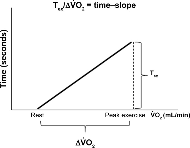
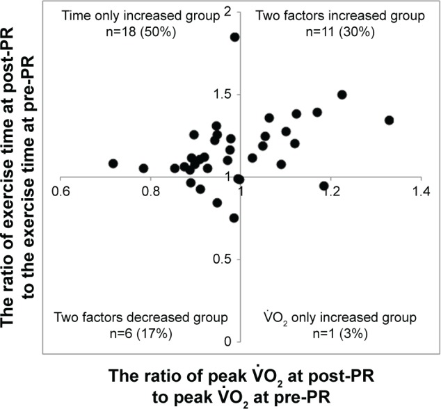
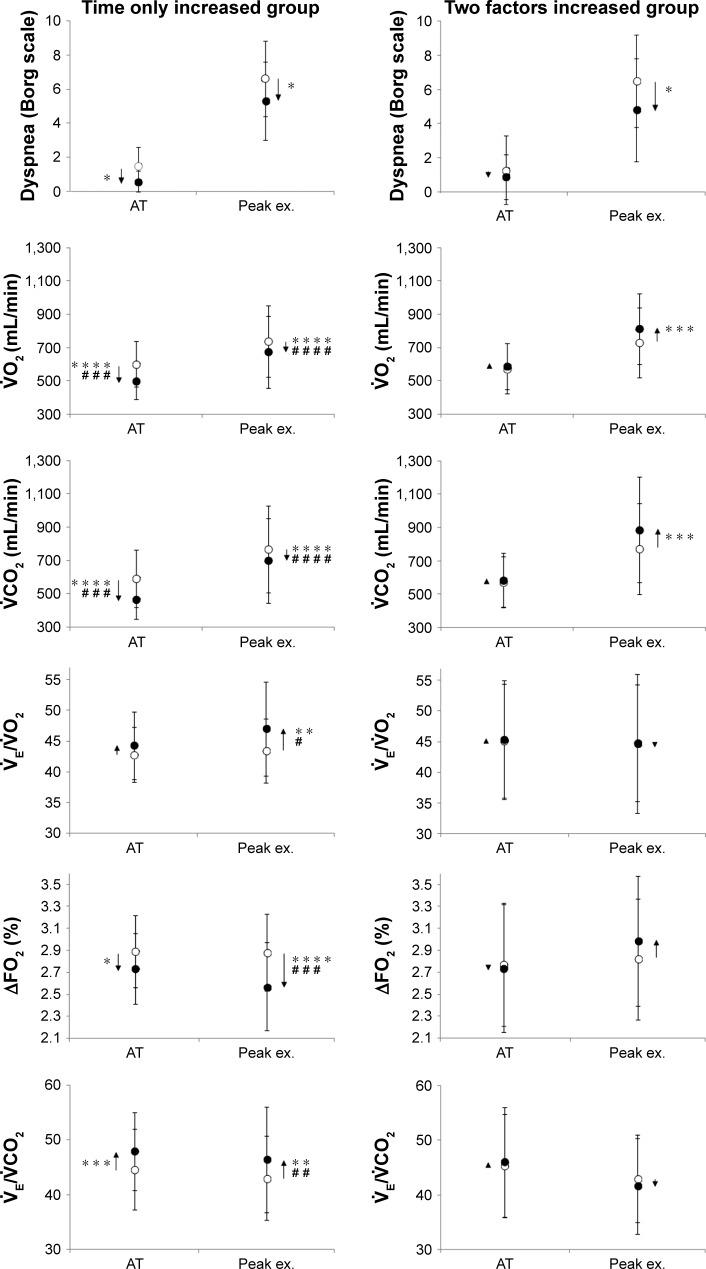
Thirty-six stable COPD patients underwent PR including a 4-week high-intensity exercise training program, and they were evaluated by cardiopulmonary exercise testing. All patients (mean age 69 years, severe and very severe COPD 94%) were classified into four groups by whether the exercise time (T) or the peak oxygen uptake [Formula: see text] increased after PR: two factors increased (both the T and the peak [Formula: see text] increased); two factors decreased; time only increased (the T increased, but the peak [Formula: see text] economized); and [Formula: see text] only increased (the T decreased, but the peak [Formula: see text] increased). Within all patients, the relationships between baseline variables and the post-to-pre-change ratio of the time-slope, T/(peak minus resting [Formula: see text]), were investigated.
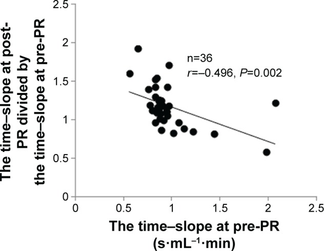
Compared with the two factors increased group (n=11), in the time only increased group (n=18), the mean differences from pre-PR at peak exercise in 1) minute ventilation [Formula: see text] (=0.004), [Formula: see text] (<0.0001), and carbon dioxide output [Formula: see text] (<0.0001) were lower, 2) [Formula: see text]/ [Formula: see text] (=0.034) and [Formula: see text]/ [Formula: see text] (=0.006) were higher, and 3) the dead space/tidal volume ratio (V/V) and the dyspnea level were similar. After PR, there was no significant difference in the ratio of the observed peak heart rate (HR) to the predicted peak HR (220 - age [years]) between the two groups. A significant negative correlation with the baseline time-slope (=-0.496, =0.002) and a positive correlation with the baseline body mass index (BMI) (=0.496, =0.002) were obtained.
PR in COPD patients improves T rather than exercise tolerance, economizing oxygen requirements, resulting in reduced ventilatory requirements without cardiac loads followed by reduced exertional dyspnea. In addition, the time-slope and BMI could be used to predict PR responses beforehand.
接受肺康复(PR)的慢性阻塞性肺疾病(COPD)患者表现出不同的反应。本研究的目的是探讨COPD患者对PR反应的可能机制和预测因素。
36例稳定期COPD患者接受了包括为期4周的高强度运动训练计划的PR,并通过心肺运动试验进行评估。所有患者(平均年龄69岁,重度和极重度COPD患者占94%)根据PR后运动时间(T)或峰值摄氧量[公式:见原文]是否增加分为四组:两个因素均增加(T和峰值[公式:见原文]均增加);两个因素均降低;仅运动时间增加(T增加,但峰值[公式:见原文]节省);以及仅峰值[公式:见原文]增加(T降低,但峰值[公式:见原文]增加)。在所有患者中,研究了基线变量与时间斜率的前后变化比值T/(峰值减去静息[公式:见原文])之间的关系。
与两个因素均增加组(n = 11)相比,仅运动时间增加组(n = 18)在运动峰值时与PR前相比的平均差异为:1)分钟通气量[公式:见原文](= 0.004)、[公式:见原文](< 0.0001)和二氧化碳排出量[公式:见原文](< 0.0001)较低;2)[公式:见原文]/[公式:见原文](= ???)和[公式:见原文]/[公式:见原文](= 0.006)较高;3)死腔/潮气量比值(V/V)和呼吸困难程度相似。PR后,两组之间观察到的峰值心率(HR)与预测的峰值HR(220 - 年龄[岁])的比值无显著差异。与基线时间斜率呈显著负相关(= -0.496,= 0.002),与基线体重指数(BMI)呈正相关(= 0.496,= 0.002)。
COPD患者的PR改善了运动时间T而非运动耐量,节省了氧气需求,导致通气需求减少且无心脏负荷,随后运动性呼吸困难减轻。此外,时间斜率和BMI可用于预先预测PR反应。