Quang Tri T, Kim Hye-Yeong, Bao Forrest Sheng, Papay Francis A, Edwards W Barry, Liu Yang
Department of Biomedical Engineering, the University of Akron, Akron, Ohio, United States of America.
Department of Radiology, University of Pittsburgh, Pittsburgh, Pennsylvania, United States of America.
PLoS One. 2017 Apr 24;12(4):e0174928. doi: 10.1371/journal.pone.0174928. eCollection 2017.
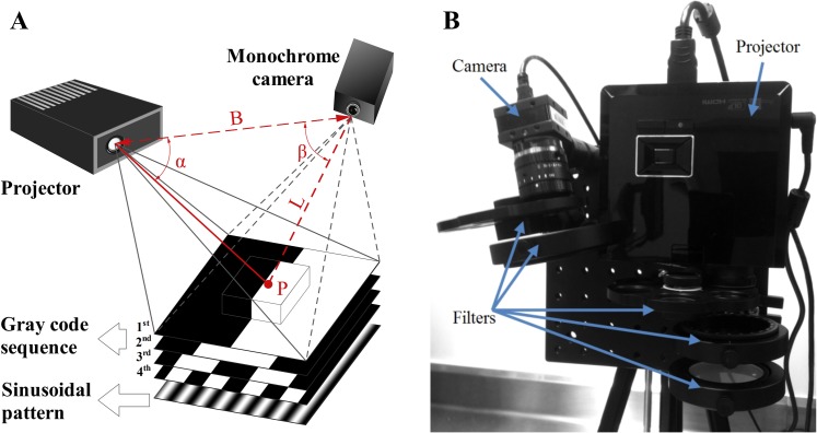
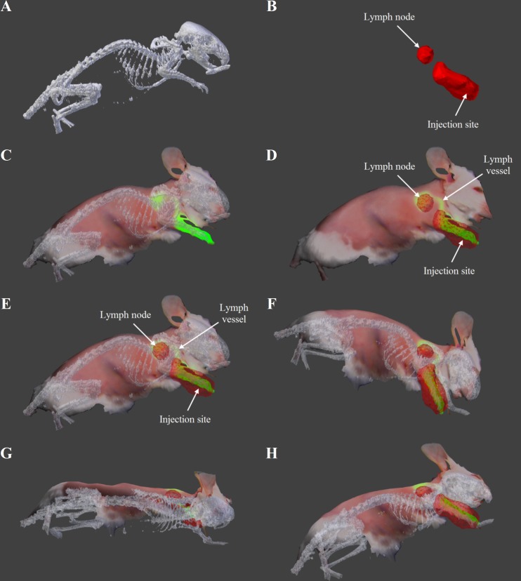
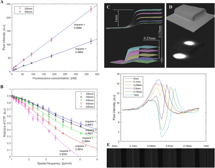
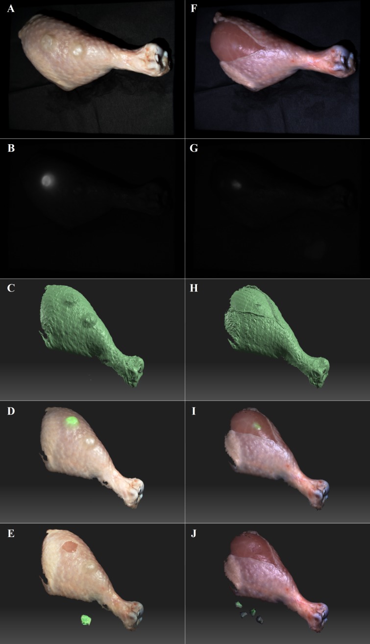
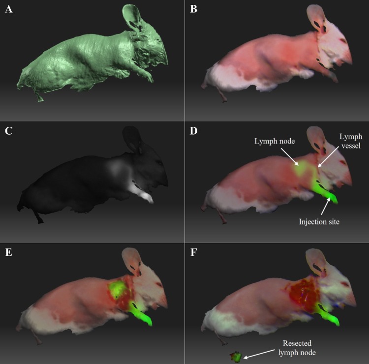
Fluorescence imaging is a powerful technique with diverse applications in intraoperative settings. Visualization of three dimensional (3D) structures and depth assessment of lesions, however, are oftentimes limited in planar fluorescence imaging systems. In this study, a novel Fluorescence Imaging Topography Scanning (FITS) system has been developed, which offers color reflectance imaging, fluorescence imaging and surface topography scanning capabilities. The system is compact and portable, and thus suitable for deployment in the operating room without disturbing the surgical flow. For system performance, parameters including near infrared fluorescence detection limit, contrast transfer functions and topography depth resolution were characterized. The developed system was tested in chicken tissues ex vivo with simulated tumors for intraoperative imaging. We subsequently conducted in vivo multimodal imaging of sentinel lymph nodes in mice using FITS and PET/CT. The PET/CT/optical multimodal images were co-registered and conveniently presented to users to guide surgeries. Our results show that the developed system can facilitate multimodal intraoperative imaging.
荧光成像技术强大,在术中环境中有多种应用。然而,在平面荧光成像系统中,三维(3D)结构的可视化以及病变深度评估常常受到限制。在本研究中,开发了一种新型荧光成像地形扫描(FITS)系统,该系统具备彩色反射成像、荧光成像和表面地形扫描功能。该系统紧凑便携,因此适合在手术室中部署,且不会干扰手术流程。针对系统性能,对包括近红外荧光检测限、对比度传递函数和地形深度分辨率等参数进行了表征。所开发的系统在离体鸡组织中使用模拟肿瘤进行了术中成像测试。随后,我们使用FITS和PET/CT对小鼠前哨淋巴结进行了体内多模态成像。PET/CT/光学多模态图像进行了配准,并方便地呈现给用户以指导手术。我们的结果表明,所开发的系统能够促进术中多模态成像。