Chi Chongwei, Du Yang, Ye Jinzuo, Kou Deqiang, Qiu Jingdan, Wang Jiandong, Tian Jie, Chen Xiaoyuan
1. Key Laboratory of Molecular Imaging of Chinese Academy of Sciences, Institute of Automation, Chinese Academy of Sciences, Beijing, 100190, China.
3. Department of General Surgery, General Hospital of People's Liberation Army, Beijing 100853, China.
Theranostics. 2014 Aug 15;4(11):1072-84. doi: 10.7150/thno.9899. eCollection 2014.
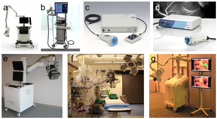
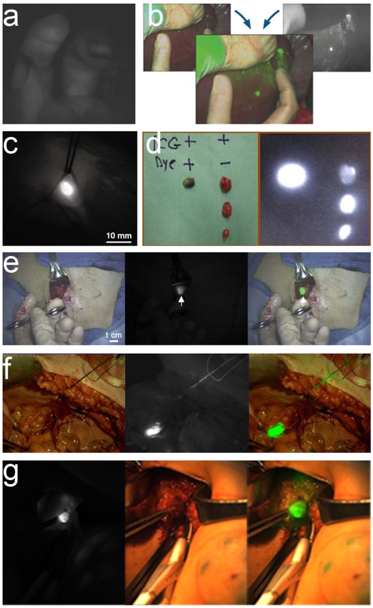
Cancer is a major threat to human health. Diagnosis and treatment using precision medicine is expected to be an effective method for preventing the initiation and progression of cancer. Although anatomical and functional imaging techniques such as radiography, computed tomography (CT), magnetic resonance imaging (MRI) and positron emission tomography (PET) have played an important role for accurate preoperative diagnostics, for the most part these techniques cannot be applied intraoperatively. Optical molecular imaging is a promising technique that provides a high degree of sensitivity and specificity in tumor margin detection. Furthermore, existing clinical applications have proven that optical molecular imaging is a powerful intraoperative tool for guiding surgeons performing precision procedures, thus enabling radical resection and improved survival rates. However, detection depth limitation exists in optical molecular imaging methods and further breakthroughs from optical to multi-modality intraoperative imaging methods are needed to develop more extensive and comprehensive intraoperative applications. Here, we review the current intraoperative optical molecular imaging technologies, focusing on contrast agents and surgical navigation systems, and then discuss the future prospects of multi-modality imaging technology for intraoperative imaging-guided cancer surgery.
癌症是对人类健康的重大威胁。使用精准医学进行诊断和治疗有望成为预防癌症发生和发展的有效方法。尽管诸如X射线摄影、计算机断层扫描(CT)、磁共振成像(MRI)和正电子发射断层扫描(PET)等解剖学和功能成像技术在准确的术前诊断中发挥了重要作用,但在很大程度上,这些技术无法在术中应用。光学分子成像技术很有前景,它在肿瘤边缘检测中具有高度的敏感性和特异性。此外,现有的临床应用已经证明,光学分子成像技术是一种强大的术中工具,可指导外科医生进行精准手术,从而实现根治性切除并提高生存率。然而,光学分子成像方法存在检测深度限制,需要从光学成像进一步突破到多模态术中成像方法,以开发更广泛、更全面的术中应用。在此,我们综述当前的术中光学分子成像技术,重点关注造影剂和手术导航系统,然后讨论多模态成像技术在术中成像引导癌症手术中的未来前景。