Sobral-Filho R G, Brito-Silva A M, Isabelle M, Jirasek A, Lum J J, Brolo A G
Department of Chemistry , University of Victoria , 3800 Finnerty Road , Victoria BC V8P 5C2 , Canada . Email:
British Columbia Cancer Agency - Vancouver Island Centre , Trev and Joyce Deeley Research Centre , 2410 Lee Ave. , Victoria , BC V8R 6V5 , Canada.
Chem Sci. 2017 Apr 1;8(4):3038-3046. doi: 10.1039/c6sc04127b. Epub 2017 Feb 3.
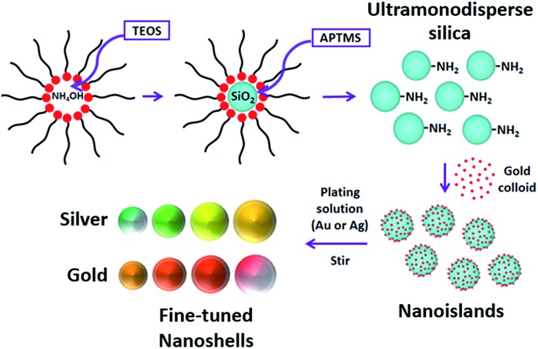
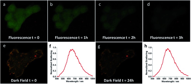
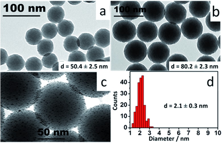
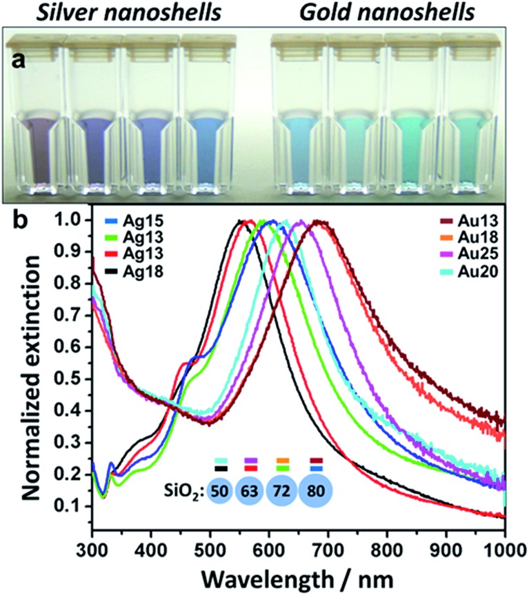
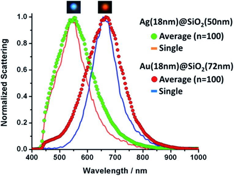
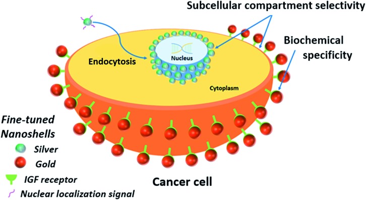
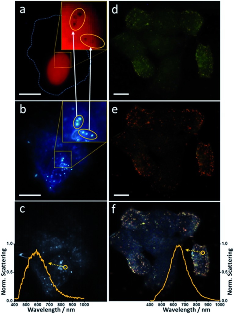
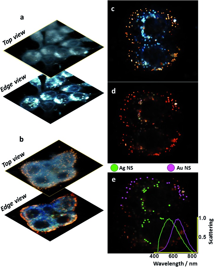
Fine-tuned gold and silver nanoshells were produced an entirely reformulated synthesis. The new method yielded ultramonodisperse samples, with polydispersity indexes (PI) as low as 0.02 and narrow extinction bands suited for multiplex analysis. A library of nanoshell samples with localized surface plasmon resonances (LSPR) spanning across the visible range was synthesized. Hyperspectral analysis revealed that the average scattering spectrum of 100 nanoshells matched closely to the spectrum of a single nanoshell, indicating an unprecedented low level of nanoparticle-to-nanoparticle variation for this type of system. A cell labeling experiment, targeting different subcellular compartments in MCF-7 human breast cancer cells, demonstrated that these monodisperse nanoparticles can be used as a multiplex platform for single cell analysis at the intracellular and extracellular level. Antibody-coated gold nanoshells targeted the plasma membrane, while silver nanoshells coated with a nuclear localization signal (NLS) targeted the nuclear membrane. A fluorescence counterstaining experiment, as well as single cell hyperspectral microscopy showed the excellent selectivity and specificity of each type of nanoparticle for its designed subcellular compartment. A time-lapse photodegradation experiment confirmed the enhanced stability of the nanoshells over fluorescent labeling and their capabilities for long-term live cell imaging.
通过全新配方的合成方法制备了经过微调的金银纳米壳。新方法得到了超单分散样品,其多分散指数(PI)低至0.02,且具有适合多重分析的窄消光带。合成了一系列具有跨越可见光范围的局域表面等离子体共振(LSPR)的纳米壳样品。高光谱分析表明,100个纳米壳的平均散射光谱与单个纳米壳的光谱非常匹配,这表明对于这类系统,纳米颗粒之间的差异达到了前所未有的低水平。针对MCF-7人乳腺癌细胞中不同亚细胞区室的细胞标记实验表明,这些单分散纳米颗粒可作为细胞内和细胞外水平单细胞分析的多重平台。抗体包被的金纳米壳靶向质膜,而包被有核定位信号(NLS)的银纳米壳靶向核膜。荧光复染实验以及单细胞高光谱显微镜显示了每种类型的纳米颗粒对其设计的亚细胞区室具有出色的选择性和特异性。时间推移光降解实验证实了纳米壳相对于荧光标记具有更高的稳定性,以及它们用于长期活细胞成像的能力。