Szobi Adrián, Gonçalvesová Eva, Varga Zoltán V, Leszek Przemyslaw, Kuśmierczyk Mariusz, Hulman Michal, Kyselovič Ján, Ferdinandy Péter, Adameová Adriana
Department of Pharmacology & Toxicology, Faculty of Pharmacy, Comenius University in Bratislava, Odbojárov 10, 832 32, Bratislava, Slovakia.
Department of Heart Failure & Transplantation, The National Institute of Cardiovascular Diseases, Bratislava, Slovakia.
J Transl Med. 2017 Apr 28;15(1):86. doi: 10.1186/s12967-017-1189-5.
Cell loss and subsequent deterioration of contractile function are hallmarks of chronic heart failure (HF). While apoptosis has been investigated as a participant in the progression of HF, it is unlikely that it accounts for the total amount of non-functional tissue. In addition, there is evidence for the presence of necrotic cardiomyocytes in HF. Therefore, the objective of this study was to investigate the necroptotic proteins regulating necroptosis, a form of programmed necrosis, and thereby assess its potential role in human end-stage HF.
Left ventricular samples of healthy controls (C) and patients with end-stage HF due to myocardial infarction (CAD) or dilated cardiomyopathy (DCM) were studied. Immunoblotting for necroptotic and apoptotic markers was performed. Triton X-114 fractionated samples were analyzed to study differences in subcellular localization.
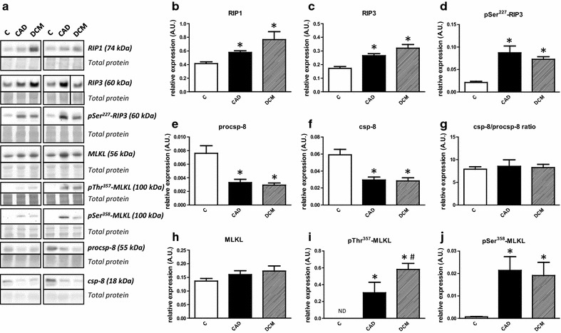
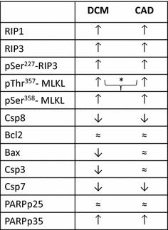
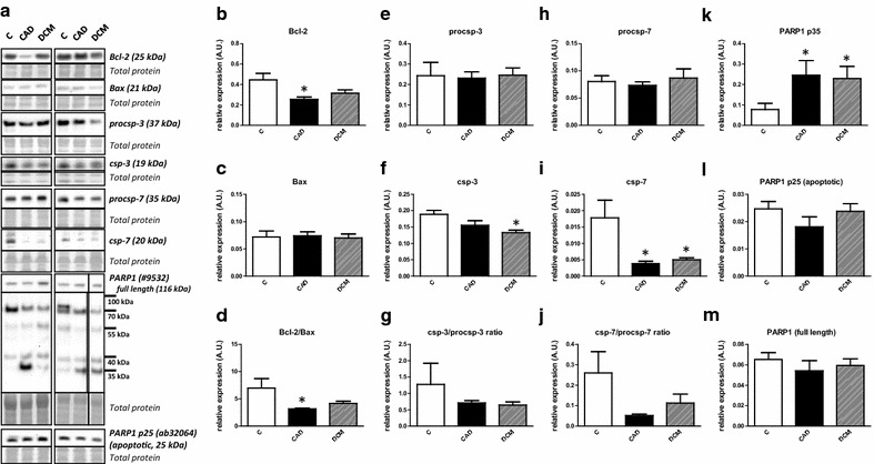
Elevated expression of RIP1 (receptor-interacting protein), pSer-RIP3 and its total levels were observed in HF groups compared to controls. On the other hand, caspase-8 expression, a proapoptotic protease negatively regulating necroptosis, was downregulated suggesting activation of necroptosis signaling. Total mixed-lineage kinase domain-like protein (MLKL) expression did not differ among the groups; however, active cytotoxic forms of MLKL were present in all HF samples while they were expressed at almost undetectable levels in controls. Interestingly, pThr-MLKL unlike pSer-MLKL, was higher in DCM than CAD. In HF, the subcellular localization of both RIP3 and pThr-MLKL was consistent with activation of necroptosis signaling. Expression of main apoptotic markers has not indicated importance of apoptosis.
This is the first evidence showing that human HF of CAD or DCM etiology is positive for markers of necroptosis which may be involved in the development of HF.
细胞丢失以及随后收缩功能的恶化是慢性心力衰竭(HF)的特征。虽然凋亡已被研究作为HF进展的参与者,但它不太可能解释无功能组织的总量。此外,有证据表明HF中存在坏死性心肌细胞。因此,本研究的目的是研究调节坏死性凋亡(一种程序性坏死形式)的坏死性凋亡蛋白,从而评估其在人类终末期HF中的潜在作用。
研究了健康对照(C)以及因心肌梗死(CAD)或扩张型心肌病(DCM)导致的终末期HF患者的左心室样本。进行了坏死性凋亡和凋亡标志物的免疫印迹分析。对经 Triton X - 114分级分离的样本进行分析,以研究亚细胞定位的差异。
与对照组相比,HF组中RIP1(受体相互作用蛋白)、pSer - RIP3及其总水平的表达升高。另一方面,半胱天冬酶 - 8的表达下调,半胱天冬酶 - 8是一种负向调节坏死性凋亡的促凋亡蛋白酶,提示坏死性凋亡信号通路被激活。各组之间总混合谱系激酶结构域样蛋白(MLKL)的表达无差异;然而,活性细胞毒性形式的MLKL存在于所有HF样本中,而在对照组中其表达水平几乎检测不到。有趣地是,与pSer - MLKL不同,pThr - MLKL在DCM中比CAD中更高。在HF中,RIP3和pThr - MLKL的亚细胞定位与坏死性凋亡信号通路的激活一致。主要凋亡标志物的表达并未表明凋亡的重要性。
这是首个证据表明,由CAD或DCM病因引起的人类HF中坏死性凋亡标志物呈阳性,这可能参与了HF的发生发展。