Domazetovic Vladana, Fontani Filippo, Marcucci Gemma, Iantomasi Teresa, Brandi Maria Luisa, Vincenzini Maria Teresa
Department of Biomedical, Experimental and Clinical Sciences "Mario Serio" (Biochemistry section) University of Florence Italy.
Department of Surgery and Translational Medicine (Endocrinology Section) University of Florence Italy.
FEBS Open Bio. 2017 Apr 5;7(5):705-718. doi: 10.1002/2211-5463.12216. eCollection 2017 May.
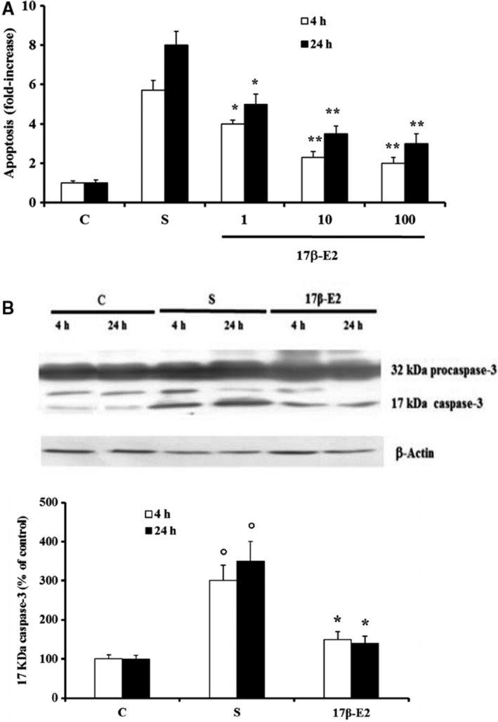
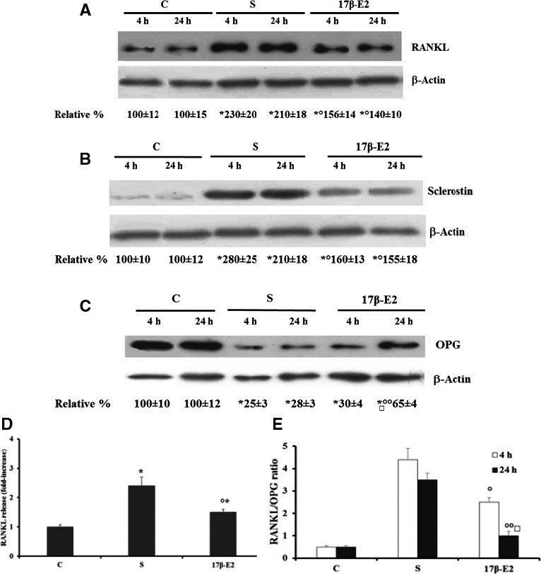
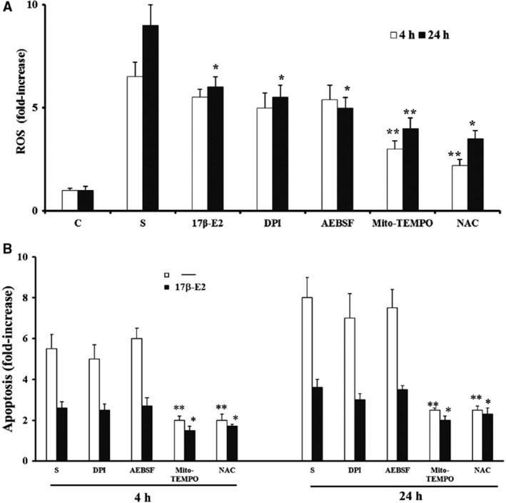
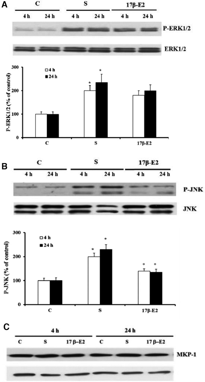
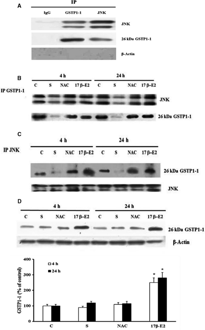
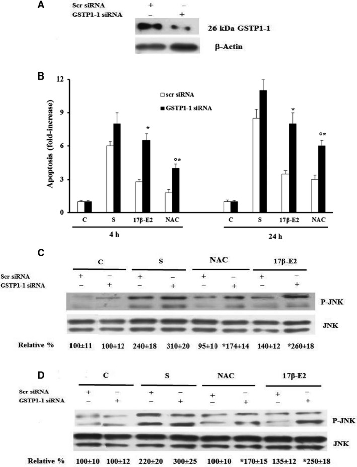
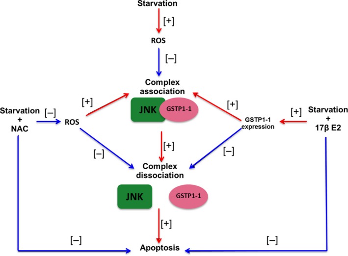
Estrogen deficiency causes bone loss as a result of microdamage, oxidative stress, and osteocyte apoptosis. A relationship between oxidative stress-induced apoptosis, c-Jun N-terminal kinase (JNK) activation, and expression of factors involved in bone remodeling has been demonstrated in osteocytes. However, the molecular regulation of these events in osteocytes treated with 17β-estradiol (17β-E2) remains unexplored. The MLO-Y4 murine osteocyte-like cell line was used as a model to study starvation-induced apoptosis and ROS production during 17β-E2 treatment. Expression of glutathione S-transferase P1-1 (GSTP1-1), receptor activator kB ligand (RANKL), osteoprotegerin (OPG), sclerostin, and kinases activation were measured by western blot. In addition, the GSTP1-1/JNK association was assessed by immunoprecipitation, and GSTP1-1 involvement in the osteocyte response to 17β-E2 was detected by specific siRNA transfection. 17β-E2 prevents starvation-induced apoptosis (DNA fragmentation and caspase activation), the increase in sclerostin expression and the RANKL/OPG ratio, which are all related to JNK activation due to oxidative stress in osteocytes. This occurs through GSTP1-1 overexpression, which can inhibit JNK activation by formation of a GSTP1-1/JNK complex. No early antioxidant action of 17β-E2 has been found but the estrogen effect is similar to N-acetylcysteine which, by increasing the intracellular redox state, maintains JNK bound to GSTP1-1. Thus, the antiapoptotic and osteogenic effect of 17β-E2 in MLO-Y4 occurs by a redox-independent process involving GSTP1-1/JNK association. This study clarifies at molecular level the effect of 17β-E2 on osteocyte activity and identifies a possible role of GSTP1-1 and JNK activity in bone remodeling and repair mechanisms.
雌激素缺乏会因微损伤、氧化应激和骨细胞凋亡导致骨质流失。在骨细胞中,氧化应激诱导的凋亡、c-Jun氨基末端激酶(JNK)激活与参与骨重塑的因子表达之间的关系已得到证实。然而,用17β-雌二醇(17β-E2)处理的骨细胞中这些事件的分子调控仍未得到探索。MLO-Y4小鼠骨细胞样细胞系被用作模型,以研究17β-E2处理期间饥饿诱导的凋亡和活性氧(ROS)产生。通过蛋白质印迹法测量谷胱甘肽S-转移酶P1-1(GSTP1-1)、核因子κB受体活化因子配体(RANKL)、骨保护素(OPG)、硬化蛋白的表达以及激酶激活情况。此外,通过免疫沉淀评估GSTP1-1/JNK的结合,并通过特异性小干扰RNA转染检测GSTP1-1参与骨细胞对17β-E2的反应。17β-E2可预防饥饿诱导的凋亡(DNA片段化和半胱天冬酶激活)、硬化蛋白表达增加以及RANKL/OPG比值升高,这些均与骨细胞中氧化应激导致的JNK激活有关。这是通过GSTP1-1的过表达实现的,GSTP1-1可通过形成GSTP1-1/JNK复合物来抑制JNK激活。未发现17β-E2有早期抗氧化作用,但雌激素的作用类似于N-乙酰半胱氨酸,后者通过增加细胞内氧化还原状态,使JNK与GSTP1-1结合。因此,17β-E2在MLO-Y4中的抗凋亡和成骨作用是通过涉及GSTP1-1/JNK结合的氧化还原非依赖性过程发生的。本研究在分子水平上阐明了17β-E2对骨细胞活性的影响,并确定了GSTP1-1和JNK活性在骨重塑和修复机制中的可能作用。