Wang Xi-Qiao, Song Fei, Liu Ying-Kai
Burn Centre, Ruijin Hospital, Jiaotong University Medical School, Shanghai, P.R. China.
PLoS One. 2017 May 4;12(5):e0176681. doi: 10.1371/journal.pone.0176681. eCollection 2017.
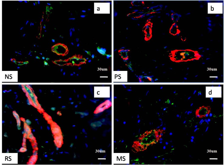
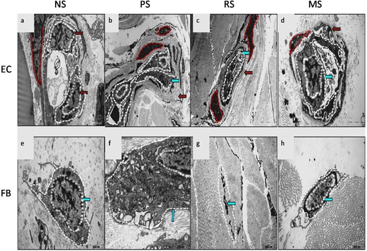
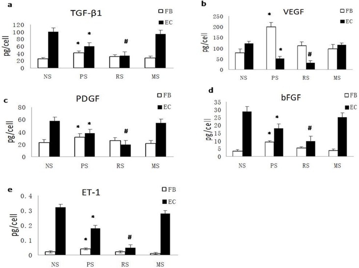
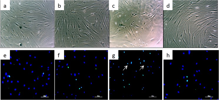
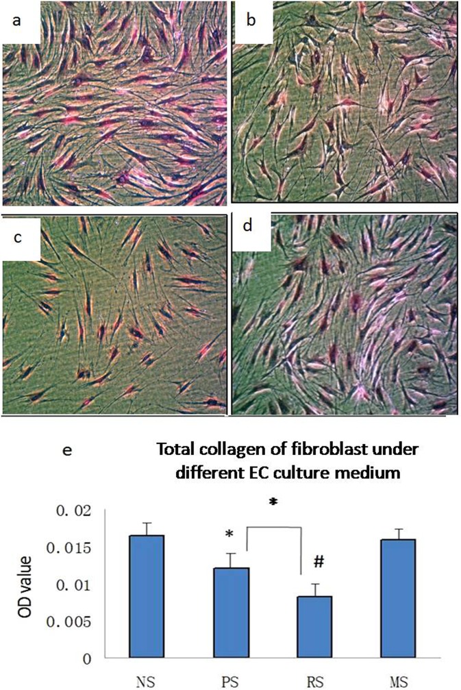
Most microvessels have been shown to become stenosed or completely occluded during hypertrophic scar progression. Here, we examined the morphology of capillary endothelial cells (ECs) and fibroblasts using immunofluorescence staining for CD31 and alpha-smooth muscle actin (α-SMA) and electron microscopy. In addition, ECs and fibroblasts were isolated from scar tissues, and the levels of transforming growth factor beta 1 (TGF-β1), platelet-derived growth factor (PDGF), endothelin 1 (ET-1), vascular endothelial growth factor (VEGF) and basic fibroblast growth factor (bFGF) were assayed using ELISAs. Furthermore, we assessed cell viability, total collagen production, and cell apoptosis in hypertrophic scar-derived fibroblasts cultured with EC-conditioned medium. Then, anti-TGF-β1, anti-PDGF, anti-ET-1, anti-VEGF, and anti-bFGF neutralising antibodies were individually added to the EC medium to identify which growth factor plays a more important role in inhibiting fibroblasts biology. Our results showed microvessel lumen occlusion and EC atrophy during scar development, particularly in regressive scars (RSs). Additionally, EC growth factor secretion decreased and reached the lowest levels in RSs. Furthermore, based on the culture results, RS EC medium inhibited fibroblast viability and collagen production and induced apoptosis. Moreover, TGF-β1, PDGF, and bFGF played more important roles in these processes than VEGF and ET-1. The endothelial dysfunction occurring in hypertrophic scars contributes to fibroblast inhibition and scar regression, and reduced TGF-β1, PDGF, and bFGF levels play key roles during this process.
多数微血管在肥厚性瘢痕进展过程中会出现狭窄或完全闭塞。在此,我们通过对CD31和α平滑肌肌动蛋白(α-SMA)进行免疫荧光染色以及电子显微镜检查,研究了毛细血管内皮细胞(ECs)和成纤维细胞的形态。此外,从瘢痕组织中分离出ECs和成纤维细胞,并使用酶联免疫吸附测定法(ELISAs)检测转化生长因子β1(TGF-β1)、血小板衍生生长因子(PDGF)、内皮素1(ET-1)、血管内皮生长因子(VEGF)和碱性成纤维细胞生长因子(bFGF)的水平。此外,我们评估了用EC条件培养基培养的肥厚性瘢痕来源的成纤维细胞的细胞活力、总胶原蛋白生成和细胞凋亡。然后,将抗TGF-β1、抗PDGF、抗ET-1、抗VEGF和抗bFGF中和抗体分别添加到EC培养基中,以确定哪种生长因子在抑制成纤维细胞生物学特性方面发挥更重要的作用。我们的结果显示,在瘢痕发展过程中,尤其是在退行性瘢痕(RSs)中,微血管腔闭塞和EC萎缩。此外,EC生长因子分泌减少,在RSs中达到最低水平。此外,根据培养结果,RS EC培养基抑制成纤维细胞活力和胶原蛋白生成并诱导细胞凋亡。此外,TGF-β1、PDGF和bFGF在这些过程中比VEGF和ET-1发挥更重要的作用。肥厚性瘢痕中发生的内皮功能障碍有助于抑制成纤维细胞和瘢痕消退,在此过程中,TGF-β1、PDGF和bFGF水平降低起关键作用。