• 文献检索
  • 文档翻译
  • 深度研究
  • 学术资讯
  • Suppr Zotero 插件Zotero 插件
  • 邀请有礼
  • 套餐&价格
  • 历史记录
应用&插件
Suppr Zotero 插件Zotero 插件浏览器插件Mac 客户端Windows 客户端微信小程序
定价
高级版会员购买积分包购买API积分包
服务
文献检索文档翻译深度研究API 文档MCP 服务
关于我们
关于 Suppr公司介绍联系我们用户协议隐私条款
关注我们

Suppr 超能文献

核心技术专利:CN118964589B侵权必究
粤ICP备2023148730 号-1Suppr @ 2026

文献检索

告别复杂PubMed语法,用中文像聊天一样搜索,搜遍4000万医学文献。AI智能推荐,让科研检索更轻松。

立即免费搜索

文件翻译

保留排版,准确专业,支持PDF/Word/PPT等文件格式,支持 12+语言互译。

免费翻译文档

深度研究

AI帮你快速写综述,25分钟生成高质量综述,智能提取关键信息,辅助科研写作。

立即免费体验

鼻腭管的评估:一项解剖学研究。

Assessment of the Nasopalatine Canal: an Anatomical Study.

作者信息

Nasseh Ibrahim, Aoun Georges, Sokhn Sayde

机构信息

Department of Dentomaxillofacial Radiology and Imaging, Faculty of Dentistry, Lebanese University, Beirut, Lebanon.

Department of Oral Pathology and Diagnosis, Faculty of Dentistry, Lebanese University, Beirut, Lebanon.

出版信息

Acta Inform Med. 2017 Mar;25(1):34-38. doi: 10.5455/aim.2017.25.34-38.

DOI:10.5455/aim.2017.25.34-38
PMID:28484295
原文链接:https://pmc.ncbi.nlm.nih.gov/articles/PMC5402381/
Abstract

OBJECTIVE

The aim of this study was to evaluate the anatomy of the nasopalatine canal in a Lebanese population using cone-beam computed tomography (CBCT) technology.

MATERIALS AND METHODS

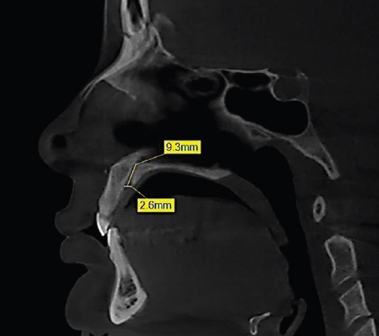
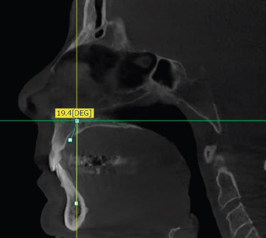
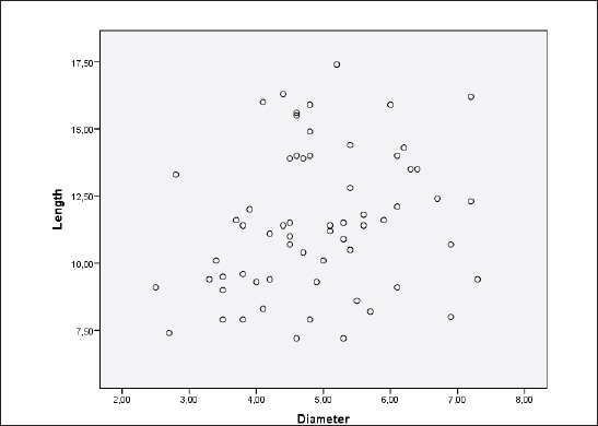
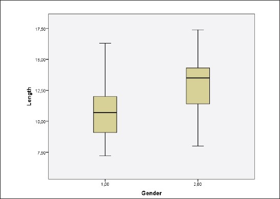
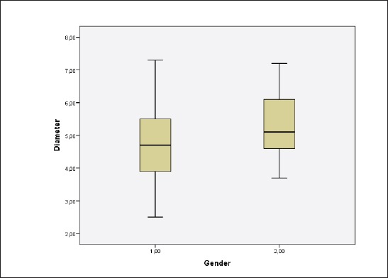
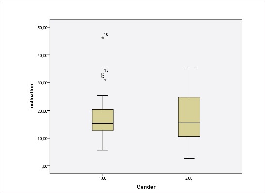
CBCT images of 63 Lebanese adult patients were included in this study. The length, shape, diameter of the oral opening corresponding to the incisive foramen and inclination in relation to the hard palate of the nasopalatine canal were analyzed.

RESULTS

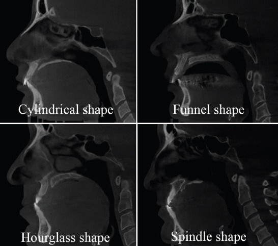
Of all canals assessed, 13 were hourglass-shaped, 23 were cylindrical-shaped, 23 were funnel-shaped and 4 were spindle-shaped. The mean canal length and the incisive foramen anteroposterior diameter were respectively 11.52 mm and 4.91 mm. The average canal inclination related to the hard palate was 17.09 degrees.

CONCLUSION

Within the limits of this study, we conclude that in Lebanese patients, the shape of the nasopalatine canal is variable. No statistical significance was noticed between genders except for the canal length which was found shorter in our female sample.

摘要

目的

本研究旨在利用锥形束计算机断层扫描(CBCT)技术评估黎巴嫩人群中鼻腭管的解剖结构。

材料与方法

本研究纳入了63名黎巴嫩成年患者的CBCT图像。分析了与切牙孔对应的口腔开口的长度、形状、直径以及鼻腭管相对于硬腭的倾斜度。

结果

在所有评估的管道中,13个呈沙漏形,23个呈圆柱形,23个呈漏斗形,4个呈纺锤形。平均管道长度和切牙孔前后径分别为11.52毫米和4.91毫米。管道相对于硬腭的平均倾斜度为17.09度。

结论

在本研究的范围内,我们得出结论,在黎巴嫩患者中,鼻腭管的形状是可变的。除了在我们的女性样本中发现管道长度较短外,未发现性别之间有统计学意义。

https://cdn.ncbi.nlm.nih.gov/pmc/blobs/753e/5402381/3426ebc2d8e5/AIM-25-34-g008.jpg
https://cdn.ncbi.nlm.nih.gov/pmc/blobs/753e/5402381/7f748a337d5b/AIM-25-34-g001.jpg
https://cdn.ncbi.nlm.nih.gov/pmc/blobs/753e/5402381/c95b0b0cdc9a/AIM-25-34-g002.jpg
https://cdn.ncbi.nlm.nih.gov/pmc/blobs/753e/5402381/82b91659ae9d/AIM-25-34-g003.jpg
https://cdn.ncbi.nlm.nih.gov/pmc/blobs/753e/5402381/7aa714d4c445/AIM-25-34-g004.jpg
https://cdn.ncbi.nlm.nih.gov/pmc/blobs/753e/5402381/b56ed9abc691/AIM-25-34-g005.jpg
https://cdn.ncbi.nlm.nih.gov/pmc/blobs/753e/5402381/f5e56fedf627/AIM-25-34-g006.jpg
https://cdn.ncbi.nlm.nih.gov/pmc/blobs/753e/5402381/cbda5cc28269/AIM-25-34-g007.jpg
https://cdn.ncbi.nlm.nih.gov/pmc/blobs/753e/5402381/3426ebc2d8e5/AIM-25-34-g008.jpg

相似文献

1
Assessment of the Nasopalatine Canal: an Anatomical Study.鼻腭管的评估:一项解剖学研究。
Acta Inform Med. 2017 Mar;25(1):34-38. doi: 10.5455/aim.2017.25.34-38.
2
Anatomical evaluation of nasopalatine canal on cone beam computed tomography images.锥形束计算机断层扫描图像上鼻腭管的解剖学评估
Folia Morphol (Warsz). 2019;78(1):153-162. doi: 10.5603/FM.a2018.0062. Epub 2018 Jul 16.
3
Morphological Variations of the Nasopalatine Canal in the North Cyprus Population: A Cone Beam Computed Tomography Study.北塞浦路斯人群鼻腭管的形态学变异:锥形束 CT 研究。
Med Sci Monit. 2024 Jun 29;30:e944868. doi: 10.12659/MSM.944868.
4
Morphometric analysis of nasopalatine foramen in Sri Lankan population using CBCT.使用锥形束计算机断层扫描(CBCT)对斯里兰卡人群鼻腭孔进行形态测量分析。
J Oral Biol Craniofac Res. 2020 Apr-Jun;10(2):238-240. doi: 10.1016/j.jobcr.2019.11.002. Epub 2019 Dec 3.
5
Morphological evaluation of the nasopalatine canal using cone beam computed tomography and its clinical implications for orthodontic miniscrew insertion.应用锥形束 CT 对鼻腭管进行形态学评估及其在正畸微种植钉植入中的临床意义。
Dent Med Probl. 2024 May-Jun;61(3):363-371. doi: 10.17219/dmp/159154.
6
Anatomical and Morphological Assessment of Nasopalatine Canal in Pediatric and Adolescent Population via Cone Beam Computed Tomography.经锥形束计算机断层扫描评估儿科和青少年人群的鼻腭管的解剖和形态。
J Craniofac Surg. 2021 Sep 1;32(6):1994-1998. doi: 10.1097/SCS.0000000000007476.
7
Morphological CBCT parameters for an accurate differentiation between nasopalatine duct cyst and the normal nasopalatine canal.用于准确区分鼻腭管囊肿和正常鼻腭管的形态 CBCT 参数。
Head Face Med. 2024 Sep 28;20(1):54. doi: 10.1186/s13005-024-00458-6.
8
Evaluation of Morphology and Anatomical Measurement of Nasopalatine Canal Using Cone Beam Computed Tomography.使用锥形束计算机断层扫描评估鼻腭管的形态和解剖测量
J Dent (Tehran). 2016 Aug;13(4):287-294.
9
The Nasopalatine Canal in Adults on Cone Beam Computed Tomograms-A Clinical Study and Review of the Literature.锥形束计算机断层扫描下成人的鼻腭管——一项临床研究及文献综述
In Vivo. 2015 Jul-Aug;29(4):467-86.
10
Cone beam computed tomography assessment of the maxillary incisive canal and foramen: considerations of anatomical variations when placing immediate implants.锥形束计算机断层扫描对上颌切牙管和切牙孔的评估:即刻种植时解剖变异的考量
PLoS One. 2015 Feb 13;10(2):e0117251. doi: 10.1371/journal.pone.0117251. eCollection 2015.

引用本文的文献

1
Morphology and dimensions of the nasopalatine canal in Palestinian patients assessed using cone beam computed tomography.使用锥形束计算机断层扫描评估巴勒斯坦患者鼻腭管的形态和尺寸。
Sci Rep. 2025 Apr 17;15(1):13248. doi: 10.1038/s41598-025-97392-7.
2
Assessment of variations in the nasopalatine canal on CBCT: considerations from an anatomical point of view.锥形束CT(CBCT)对鼻腭管变异的评估:解剖学角度的考量
J Periodontal Implant Sci. 2025 Feb;55(1):62-71. doi: 10.5051/jpis.2401300065. Epub 2024 Jul 12.
3
Morphological Variations of the Nasopalatine Canal in the North Cyprus Population: A Cone Beam Computed Tomography Study.

本文引用的文献

1
Three-dimensional analysis of incisive canals in human dentulous and edentulous maxillary bones.人类有牙和无牙上颌骨中切牙管的三维分析
Int J Implant Dent. 2015 Dec;1(1):12. doi: 10.1186/s40729-015-0012-4. Epub 2015 May 1.
2
Evaluation of the nasopalatine canal with cone-beam computed tomography in an Iranian population.伊朗人群中锥形束计算机断层扫描对鼻腭管的评估。
Dent Res J (Isfahan). 2015 Jan-Feb;12(1):14-9. doi: 10.4103/1735-3327.150289.
3
Cone beam computed tomography assessment of the maxillary incisive canal and foramen: considerations of anatomical variations when placing immediate implants.
北塞浦路斯人群鼻腭管的形态学变异:锥形束 CT 研究。
Med Sci Monit. 2024 Jun 29;30:e944868. doi: 10.12659/MSM.944868.
4
Assessment of the Nasopalatine Canal in Patients Requiring Dental Implants in the Maxillary Anterior Region Using Cone Beam Computed Tomography.使用锥形束计算机断层扫描评估上颌前部区域需要种植牙的患者的鼻腭管。
Cureus. 2023 Dec 16;15(12):e50643. doi: 10.7759/cureus.50643. eCollection 2023 Dec.
5
Are morphological and morphometric characteristics of maxillary anterior region and nasopalatine canal related to each other?上颌前部区域和鼻腭管的形态学及形态测量学特征彼此相关吗?
Oral Radiol. 2023 Apr;39(2):372-385. doi: 10.1007/s11282-022-00647-6. Epub 2022 Aug 8.
6
Evaluation of Buccal Bone Wall Thickness of Anterosuperior Teeth and Nasopalatine Duct Morphology in Cone Beam Computed Tomography of Patients Living at Different Altitudes: A Two-Year Retrospective Study.不同海拔地区患者锥形束计算机断层扫描中上前牙颊侧骨壁厚度及鼻腭管形态的评估:一项为期两年的回顾性研究
J Int Soc Prev Community Dent. 2021 Nov 30;11(6):652-660. doi: 10.4103/jispcd.JISPCD_126_21. eCollection 2021 Nov-Dec.
7
Morphological Characteristics of the Nasopalatine Canal and the Relationship with the Anterior Maxillary Bone-A Cone Beam Computed Tomography Study.鼻腭管的形态学特征及其与上颌前部骨的关系——锥形束计算机断层扫描研究
Diagnostics (Basel). 2021 May 20;11(5):915. doi: 10.3390/diagnostics11050915.
8
Morphometric analysis of nasopalatine foramen in Sri Lankan population using CBCT.使用锥形束计算机断层扫描(CBCT)对斯里兰卡人群鼻腭孔进行形态测量分析。
J Oral Biol Craniofac Res. 2020 Apr-Jun;10(2):238-240. doi: 10.1016/j.jobcr.2019.11.002. Epub 2019 Dec 3.
9
Cone beam computerized tomography evaluation of incisive canal and anterior maxillary bone thickness for placement of immediate implants.锥形束计算机断层扫描评估切牙管及上颌前部骨厚度以用于即刻种植体植入
J Indian Prosthodont Soc. 2018 Oct-Dec;18(4):356-363. doi: 10.4103/jips.jips_167_18.
10
The Incisive Canal: A Comprehensive Review.切牙管:全面综述
Cureus. 2018 Jul 30;10(7):e3069. doi: 10.7759/cureus.3069.
锥形束计算机断层扫描对上颌切牙管和切牙孔的评估:即刻种植时解剖变异的考量
PLoS One. 2015 Feb 13;10(2):e0117251. doi: 10.1371/journal.pone.0117251. eCollection 2015.
4
Anatomy and morphology of the nasopalatine canal using cone-beam computed tomography.使用锥形束计算机断层扫描技术观察鼻腭管的解剖结构和形态
Imaging Sci Dent. 2013 Dec;43(4):273-81. doi: 10.5624/isd.2013.43.4.273. Epub 2013 Dec 12.
5
Evaluation of maxillary incisive canal characteristics related to dental implant treatment with computerized tomography: a clinical multicenter study.应用计算机断层扫描评估与牙种植治疗相关的上颌切牙管特征:一项临床多中心研究。
J Periodontol. 2012 Mar;83(3):337-43. doi: 10.1902/jop.2011.110326. Epub 2011 Sep 12.
6
Morphology of the nasopalatine canal and dental implant surgery: a radiographic analysis of 100 consecutive patients using limited cone-beam computed tomography.应用锥形束 CT 对 100 例患者的鼻腭管形态及牙种植手术的放射学分析
Clin Oral Implants Res. 2011 Mar;22(3):295-301. doi: 10.1111/j.1600-0501.2010.02010.x. Epub 2010 Oct 6.
7
Microanatomy of the incisive canal using three-dimensional reconstruction of microCT images: an ex vivo study.使用显微CT图像三维重建对切牙管进行显微解剖:一项离体研究
Oral Surg Oral Med Oral Pathol Oral Radiol Endod. 2009 Oct;108(4):583-90. doi: 10.1016/j.tripleo.2009.06.036.
8
Macro- and micro-anatomical, histological and computed tomography scan characterization of the nasopalatine canal.鼻腭管的大体与显微解剖、组织学及计算机断层扫描特征
J Clin Periodontol. 2009 Jul;36(7):598-603. doi: 10.1111/j.1600-051X.2009.01429.x.
9
Morphologic changes of the nasopalatine canal related to dental implantation: a radiologic study in different degrees of absorbed maxillae.与牙种植相关的鼻腭管形态学变化:不同程度上颌骨吸收的影像学研究
J Periodontol. 2008 Sep;79(9):1659-62. doi: 10.1902/jop.2008.080043.
10
The nasopalatine canal revisited using 2D and 3D CT imaging.使用二维和三维CT成像重新审视鼻腭管。
Dentomaxillofac Radiol. 2004 Nov;33(6):396-402. doi: 10.1259/dmfr/53801969.