Sözer Esin B, Pocetti C Florencia, Vernier P Thomas
Frank Reidy Research Center for Bioelectrics, Old Dominion University, 4211 Monarch Way, Ste. 300, Norfolk, VA, 23508, USA.
Department of Bioengineering, Instituto Tecnológico de Buenos Aires, Buenos Aires, Argentina.
J Membr Biol. 2018 Apr;251(2):197-210. doi: 10.1007/s00232-017-9962-1. Epub 2017 May 8.
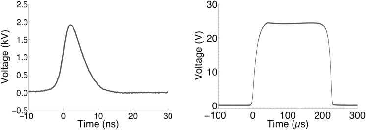
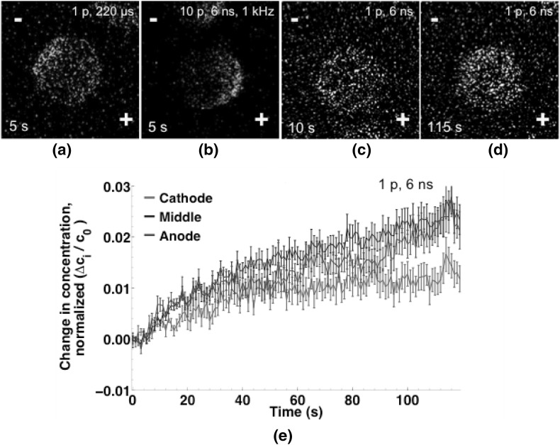
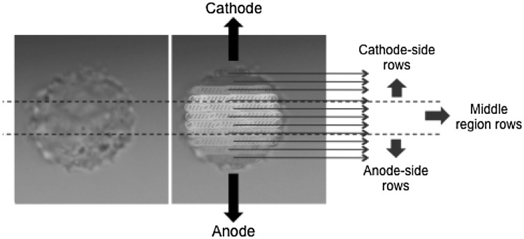
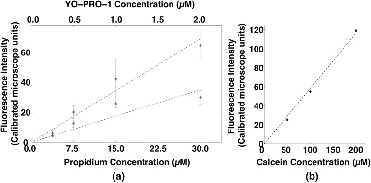
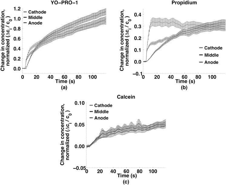
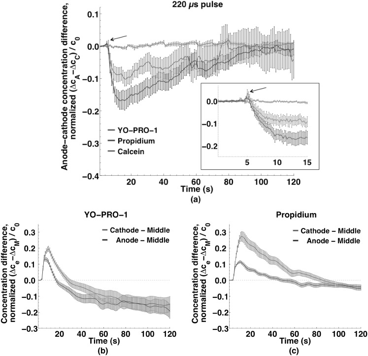
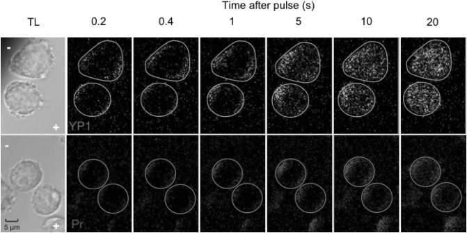
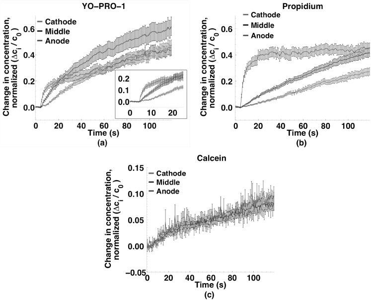
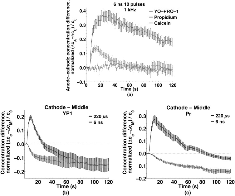
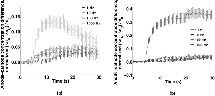
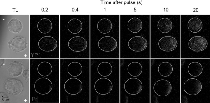
Imaging of fluorescent small molecule transport into electropermeabilized cells reveals polarized patterns of entry, which must reflect in some way the mechanisms of the migration of these molecules across the compromised membrane barrier. In some reports, transport occurs primarily across the areas of the membrane nearest the positive electrode (anode), but in others cathode-facing entry dominates. Here we compare YO-PRO-1, propidium, and calcein uptake into U-937 cells after nanosecond (6 ns) and microsecond (220 µs) electric pulse exposures. Each of the three dyes exhibits a different pattern. Calcein shows no preference for anode- or cathode-facing entry that is detectable with our measurement system. Immediately after a microsecond pulse, YO-PRO-1 and propidium enter the cell roughly equally from the positive and negative poles, but transport through the cathode-facing side dominates in less than 1 s. After nanosecond pulse permeabilization, YO-PRO-1 and propidium enter primarily on the anode-facing side of the cell.
对荧光小分子进入电穿孔细胞的成像揭示了极化的进入模式,这必定在某种程度上反映了这些分子跨越受损膜屏障的迁移机制。在一些报告中,转运主要发生在膜上最靠近正电极(阳极)的区域,但在其他报告中,面向阴极的进入占主导。在这里,我们比较了纳秒(6纳秒)和微秒(220微秒)电脉冲暴露后,YO-PRO-1、碘化丙啶和钙黄绿素进入U-937细胞的情况。这三种染料中的每一种都呈现出不同的模式。钙黄绿素对面向阳极或阴极的进入没有偏好,我们的测量系统无法检测到这种偏好。微秒脉冲后立即观察到,YO-PRO-1和碘化丙啶从正负极进入细胞的情况大致相同,但在不到1秒的时间内,通过面向阴极一侧的转运占主导。纳秒脉冲通透化后,YO-PRO-1和碘化丙啶主要在细胞面向阳极的一侧进入。