• 文献检索
  • 文档翻译
  • 深度研究
  • 学术资讯
  • Suppr Zotero 插件Zotero 插件
  • 邀请有礼
  • 套餐&价格
  • 历史记录
应用&插件
Suppr Zotero 插件Zotero 插件浏览器插件Mac 客户端Windows 客户端微信小程序
定价
高级版会员购买积分包购买API积分包
服务
文献检索文档翻译深度研究API 文档MCP 服务
关于我们
关于 Suppr公司介绍联系我们用户协议隐私条款
关注我们

Suppr 超能文献

核心技术专利:CN118964589B侵权必究
粤ICP备2023148730 号-1Suppr @ 2026

文献检索

告别复杂PubMed语法,用中文像聊天一样搜索,搜遍4000万医学文献。AI智能推荐,让科研检索更轻松。

立即免费搜索

文件翻译

保留排版,准确专业,支持PDF/Word/PPT等文件格式,支持 12+语言互译。

免费翻译文档

深度研究

AI帮你快速写综述,25分钟生成高质量综述,智能提取关键信息,辅助科研写作。

立即免费体验

在质膜复杂脂质结构中电穿孔位点的识别。

Identification of electroporation sites in the complex lipid organization of the plasma membrane.

作者信息

Rems Lea, Tang Xinru, Zhao Fangwei, Pérez-Conesa Sergio, Testa Ilaria, Delemotte Lucie

机构信息

KTH Royal Institute of Technology, Dept. Applied Physics, Science for Life Laboratory, Solna, Sweden.

University of Ljubljana, Faculty of Electrical Engineering, Ljubljana, Slovenia.

出版信息

Elife. 2022 Feb 23;11:e74773. doi: 10.7554/eLife.74773.

DOI:10.7554/eLife.74773
PMID:35195069
原文链接:https://pmc.ncbi.nlm.nih.gov/articles/PMC8912918/
Abstract

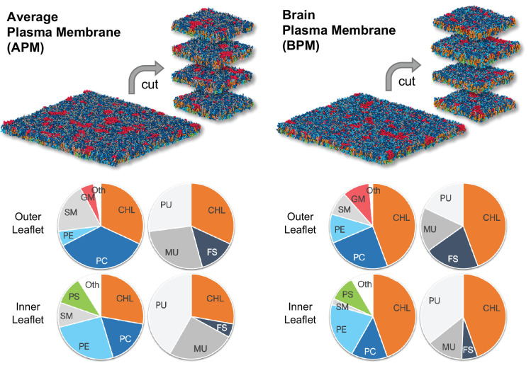
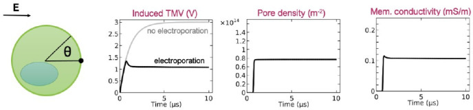
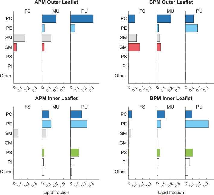
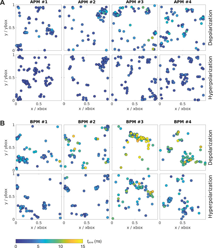
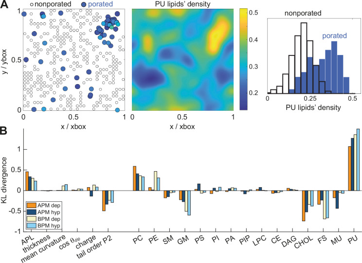
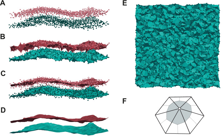
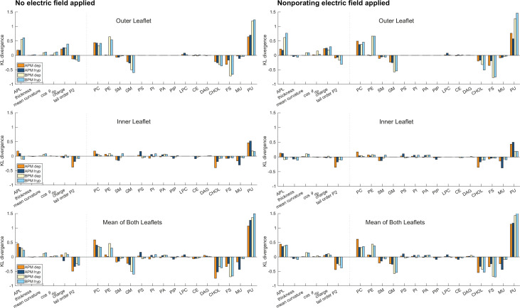
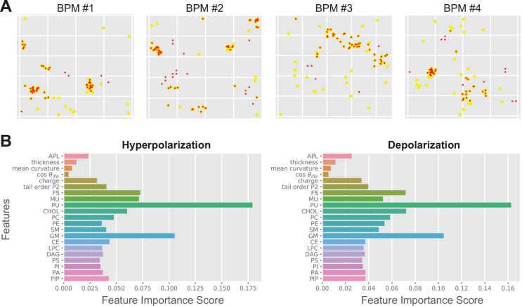
The plasma membrane of a biological cell is a complex assembly of lipids and membrane proteins, which tightly regulate transmembrane transport. When a cell is exposed to strong electric field, the membrane integrity becomes transiently disrupted by formation of transmembrane pores. This phenomenon termed electroporation is already utilized in many rapidly developing applications in medicine including gene therapy, cancer treatment, and treatment of cardiac arrhythmias. However, the molecular mechanisms of electroporation are not yet sufficiently well understood; in particular, it is unclear where exactly pores form in the complex organization of the plasma membrane. In this study, we combine coarse-grained molecular dynamics simulations, machine learning methods, and Bayesian survival analysis to identify how formation of pores depends on the local lipid organization. We show that pores do not form homogeneously across the membrane, but colocalize with domains that have specific features, the most important being high density of polyunsaturated lipids. We further show that knowing the lipid organization is sufficient to reliably predict poration sites with machine learning. Additionally, by analysing poration kinetics with Bayesian survival analysis we show that poration does not depend solely on local lipid arrangement, but also on membrane mechanical properties and the polarity of the electric field. Finally, we discuss how the combination of atomistic and coarse-grained molecular dynamics simulations, machine learning methods, and Bayesian survival analysis can guide the design of future experiments and help us to develop an accurate description of plasma membrane electroporation on the whole-cell level. Achieving this will allow us to shift the optimization of electroporation applications from blind trial-and-error approaches to mechanistic-driven design.

摘要

生物细胞的质膜是脂质和膜蛋白的复杂集合体,它严格调控跨膜运输。当细胞暴露于强电场时,跨膜孔的形成会使膜的完整性暂时遭到破坏。这种被称为电穿孔的现象已在医学领域许多快速发展的应用中得到利用,包括基因治疗、癌症治疗和心律失常治疗。然而,电穿孔的分子机制尚未得到充分理解;特别是,在质膜复杂的组织结构中,孔究竟在何处形成尚不清楚。在本研究中,我们结合粗粒度分子动力学模拟、机器学习方法和贝叶斯生存分析,以确定孔的形成如何依赖于局部脂质组织。我们发现,孔并非在整个膜上均匀形成,而是与具有特定特征的区域共定位,其中最重要的特征是多不饱和脂质的高密度。我们进一步表明,了解脂质组织足以通过机器学习可靠地预测穿孔位点。此外,通过用贝叶斯生存分析来分析穿孔动力学,我们发现穿孔不仅取决于局部脂质排列,还取决于膜的机械性能和电场的极性。最后,我们讨论了原子尺度和粗粒度分子动力学模拟、机器学习方法以及贝叶斯生存分析的结合如何能够指导未来实验的设计,并帮助我们在全细胞水平上准确描述质膜电穿孔。实现这一点将使我们能够将电穿孔应用的优化从盲目试错方法转变为机制驱动的设计。

https://cdn.ncbi.nlm.nih.gov/pmc/blobs/8019/8912918/df7a6e1b6211/elife-74773-sa2-fig2.jpg
https://cdn.ncbi.nlm.nih.gov/pmc/blobs/8019/8912918/e4c073677db6/elife-74773-fig1.jpg
https://cdn.ncbi.nlm.nih.gov/pmc/blobs/8019/8912918/2ca2fca21472/elife-74773-fig1-figsupp1.jpg
https://cdn.ncbi.nlm.nih.gov/pmc/blobs/8019/8912918/4508ad49155c/elife-74773-fig2.jpg
https://cdn.ncbi.nlm.nih.gov/pmc/blobs/8019/8912918/f4ade8488304/elife-74773-fig3.jpg
https://cdn.ncbi.nlm.nih.gov/pmc/blobs/8019/8912918/7265b20d9128/elife-74773-fig3-figsupp1.jpg
https://cdn.ncbi.nlm.nih.gov/pmc/blobs/8019/8912918/d1489bc6a602/elife-74773-fig3-figsupp2.jpg
https://cdn.ncbi.nlm.nih.gov/pmc/blobs/8019/8912918/852b4f6a37f2/elife-74773-fig4.jpg
https://cdn.ncbi.nlm.nih.gov/pmc/blobs/8019/8912918/87b029b8f758/elife-74773-fig5.jpg
https://cdn.ncbi.nlm.nih.gov/pmc/blobs/8019/8912918/816d0cf38d9f/elife-74773-fig5-figsupp1.jpg
https://cdn.ncbi.nlm.nih.gov/pmc/blobs/8019/8912918/a5f5347810a8/elife-74773-fig5-figsupp2.jpg
https://cdn.ncbi.nlm.nih.gov/pmc/blobs/8019/8912918/840ad8ab81b7/elife-74773-fig5-figsupp3.jpg
https://cdn.ncbi.nlm.nih.gov/pmc/blobs/8019/8912918/ab114b0cf72f/elife-74773-fig5-figsupp4.jpg
https://cdn.ncbi.nlm.nih.gov/pmc/blobs/8019/8912918/891d75204461/elife-74773-fig5-figsupp5.jpg
https://cdn.ncbi.nlm.nih.gov/pmc/blobs/8019/8912918/0d80594d1dfa/elife-74773-fig5-figsupp6.jpg
https://cdn.ncbi.nlm.nih.gov/pmc/blobs/8019/8912918/efa83a6b3c6d/elife-74773-sa2-fig1.jpg
https://cdn.ncbi.nlm.nih.gov/pmc/blobs/8019/8912918/df7a6e1b6211/elife-74773-sa2-fig2.jpg

相似文献

1
Identification of electroporation sites in the complex lipid organization of the plasma membrane.在质膜复杂脂质结构中电穿孔位点的识别。
Elife. 2022 Feb 23;11:e74773. doi: 10.7554/eLife.74773.
2
The importance of membrane defects-lessons from simulations.膜缺陷的重要性:模拟研究的启示。
Acc Chem Res. 2014 Aug 19;47(8):2244-51. doi: 10.1021/ar4002729. Epub 2014 Jun 3.
3
Membrane pore formation in atomistic and coarse-grained simulations.原子尺度和粗粒度模拟中的膜孔形成
Biochim Biophys Acta. 2016 Oct;1858(10):2266-2277. doi: 10.1016/j.bbamem.2015.12.031. Epub 2015 Dec 31.
4
Synergistic effect of electric field and lipid oxidation on the permeability of cell membranes.电场和脂质氧化对细胞膜通透性的协同作用。
Biochim Biophys Acta Gen Subj. 2017 Apr;1861(4):839-847. doi: 10.1016/j.bbagen.2017.01.030. Epub 2017 Jan 27.
5
Molecular dynamics simulation of reversible electroporation with Martini force field.用 Martini 力场进行可逆电穿孔的分子动力学模拟。
Biomed Eng Online. 2019 Dec 26;18(1):123. doi: 10.1186/s12938-019-0743-1.
6
Pulsed Electric Fields Can Create Pores in the Voltage Sensors of Voltage-Gated Ion Channels.脉冲电场可在电压门控离子通道的电压传感器中形成孔道。
Biophys J. 2020 Jul 7;119(1):190-205. doi: 10.1016/j.bpj.2020.05.030. Epub 2020 Jun 8.
7
Molecular dynamics insights on temperature and pressure effects on electroporation.分子动力学研究温度和压力对电穿孔的影响。
Biochim Biophys Acta Biomembr. 2022 Dec 1;1864(12):184049. doi: 10.1016/j.bbamem.2022.184049. Epub 2022 Sep 14.
8
Membrane Electroporation and Electropermeabilization: Mechanisms and Models.细胞膜电穿孔和电渗透作用:机制和模型。
Annu Rev Biophys. 2019 May 6;48:63-91. doi: 10.1146/annurev-biophys-052118-115451. Epub 2019 Feb 20.
9
Electric-driven membrane poration: A rationale for water role in the kinetics of pore formation.电驱动膜穿孔:水在孔形成动力学中的作用原理。
Bioelectrochemistry. 2022 Feb;143:107987. doi: 10.1016/j.bioelechem.2021.107987. Epub 2021 Oct 26.
10
Electric field-driven water dipoles: nanoscale architecture of electroporation.电场驱动的水分子偶极子:电穿孔的纳米结构。
PLoS One. 2013 Apr 11;8(4):e61111. doi: 10.1371/journal.pone.0061111. Print 2013.

引用本文的文献

1
Novel combination of irreversible electroporation and allogenic chimeric antigen receptor (CAR) T-cell therapy synergizes therapeutic outcomes in a preclinical human pancreatic cancer mouse model.不可逆电穿孔与同种异体嵌合抗原受体(CAR)T细胞疗法的新型联合方案在临床前人类胰腺癌小鼠模型中可增强治疗效果。
bioRxiv. 2025 Aug 12:2025.08.10.669537. doi: 10.1101/2025.08.10.669537.
2
Current Research in Drug-Free Cancer Therapies.无药癌症治疗的当前研究
Bioengineering (Basel). 2025 Mar 26;12(4):341. doi: 10.3390/bioengineering12040341.
3
How pore formation in complex biological membranes is governed by lipid composition, mechanics, and lateral sorting.

本文引用的文献

1
PyMC: a modern, and comprehensive probabilistic programming framework in Python.PyMC:Python 中一个现代且全面的概率编程框架。
PeerJ Comput Sci. 2023 Sep 1;9:e1516. doi: 10.7717/peerj-cs.1516. eCollection 2023.
2
Ablation Modalities for Therapeutic Intervention in Arrhythmia-Related Cardiovascular Disease: Focus on Electroporation.心律失常相关心血管疾病治疗干预的消融方式:聚焦电穿孔
J Clin Med. 2021 Jun 16;10(12):2657. doi: 10.3390/jcm10122657.
3
Cell death due to electroporation - A review.电穿孔导致的细胞死亡——综述
复杂生物膜中的孔形成是如何受脂质组成、力学和侧向分选作用的支配。
PNAS Nexus. 2025 Feb 21;4(3):pgaf033. doi: 10.1093/pnasnexus/pgaf033. eCollection 2025 Mar.
4
Towards electrospray-assisted production of lipid-based synthetic cell assemblies.迈向基于电喷雾辅助生产的脂质基合成细胞组件。
Soft Matter. 2025 Apr 16;21(16):2977-2985. doi: 10.1039/d4sm01284d.
5
Tissue Nanotransfection Silicon Chip and Related Electroporation-Based Technologies for In Vivo Tissue Reprogramming.用于体内组织重编程的组织纳米转染硅芯片及相关基于电穿孔的技术
Nanomaterials (Basel). 2024 Jan 19;14(2):217. doi: 10.3390/nano14020217.
6
The Importance of the Plasma Membrane in Atherogenesis.质膜在动脉粥样硬化发生中的重要性。
Membranes (Basel). 2022 Oct 24;12(11):1036. doi: 10.3390/membranes12111036.
7
Assessing membrane material properties from the response of giant unilamellar vesicles to electric fields.通过巨型单层囊泡对电场的响应来评估膜材料特性。
Adv Phys X. 2023;8(1). doi: 10.1080/23746149.2022.2125342. Epub 2022 Oct 6.
Bioelectrochemistry. 2021 Oct;141:107871. doi: 10.1016/j.bioelechem.2021.107871. Epub 2021 Jun 6.
4
To Close or to Collapse: The Role of Charges on Membrane Stability upon Pore Formation.闭合还是崩溃:膜稳定性在孔形成过程中电荷的作用。
Adv Sci (Weinh). 2021 Jun;8(11):e2004068. doi: 10.1002/advs.202004068. Epub 2021 Mar 8.
5
In Vitro Cell Selectivity of Reversible and Irreversible: Electroporation in Cardiac Tissue.在体心脏组织中可逆与不可逆电穿孔的细胞选择性。
Circ Arrhythm Electrophysiol. 2021 Apr;14(4):e008817. doi: 10.1161/CIRCEP.120.008817. Epub 2021 Mar 17.
6
Simulating realistic membrane shapes.模拟真实的膜形状。
Curr Opin Cell Biol. 2021 Aug;71:103-111. doi: 10.1016/j.ceb.2021.02.009. Epub 2021 Mar 12.
7
Dye Transport through Bilayers Agrees with Lipid Electropore Molecular Dynamics.染料通过双层膜的传输与脂质电穿孔分子动力学一致。
Biophys J. 2020 Nov 3;119(9):1724-1734. doi: 10.1016/j.bpj.2020.09.028. Epub 2020 Oct 2.
8
Actin networks regulate the cell membrane permeability during electroporation.肌动蛋白网络在电穿孔过程中调节细胞膜通透性。
Biochim Biophys Acta Biomembr. 2021 Jan 1;1863(1):183468. doi: 10.1016/j.bbamem.2020.183468. Epub 2020 Aug 31.
9
The interplay of excitation and electroporation in nanosecond pulse stimulation.纳秒脉冲刺激中的激发和电穿孔相互作用。
Bioelectrochemistry. 2020 Dec;136:107598. doi: 10.1016/j.bioelechem.2020.107598. Epub 2020 Jul 15.
10
Using Nanosecond Shocks for Cardiac Defibrillation.使用纳秒级电击进行心脏除颤。
Bioelectricity. 2019 Dec 1;1(4):240-246. doi: 10.1089/bioe.2019.0030. Epub 2019 Dec 12.