Kim Johoon, Chhour Peter, Hsu Jessica, Litt Harold I, Ferrari Victor A, Popovtzer Rachela, Cormode David P
Department of Engineering, Bar-Ilan University , Ramat Gan, 5290002, Israel.
Bioconjug Chem. 2017 Jun 21;28(6):1581-1597. doi: 10.1021/acs.bioconjchem.7b00194. Epub 2017 May 18.
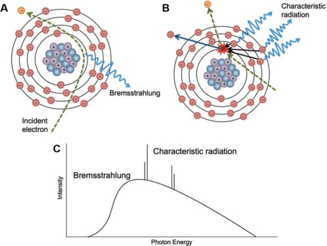
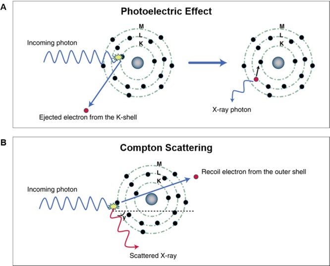
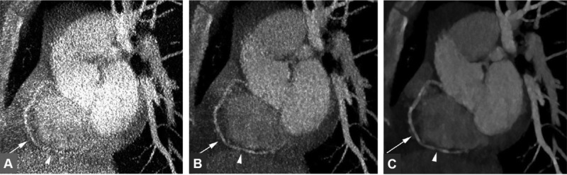
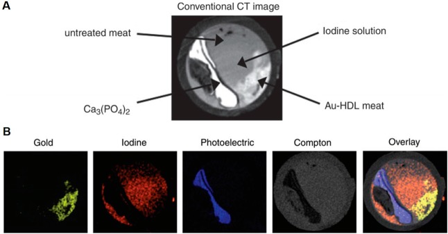
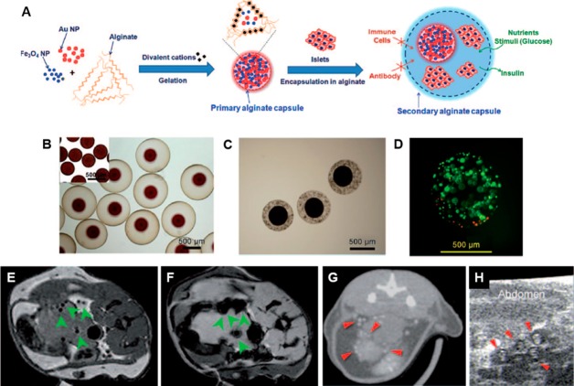
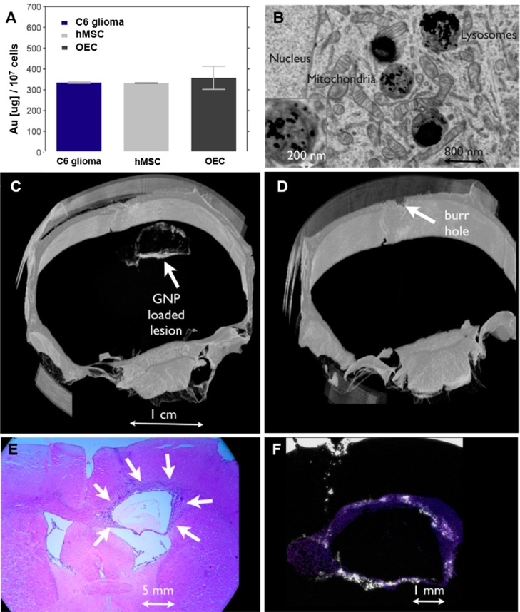
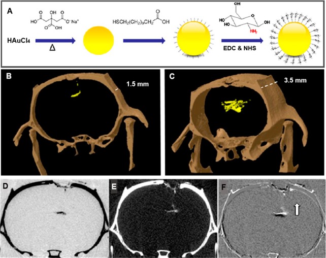
Efforts to develop novel cell-based therapies originated with the first bone marrow transplant on a leukemia patient in 1956. Preclinical and clinical examples of cell-based treatment strategies have shown promising results across many disciplines in medicine, with recent advances in immune cell therapies for cancer producing remarkable response rates, even in patients with multiple treatment failures. However, cell-based therapies suffer from inconsistent outcomes, motivating the search for tools that allow monitoring of cell delivery and behavior in vivo. Noninvasive cell imaging techniques, also known as cell tracking, have been developed to address this issue. These tools can allow real-time, quantitative, and long-term monitoring of transplanted cells in the recipient, providing insight on cell migration, distribution, viability, differentiation, and fate, all of which play crucial roles in treatment efficacy. Understanding these parameters allows the optimization of cell choice, delivery route, and dosage for therapy and advances cell-based therapy for specific clinical uses. To date, most cell tracking work has centered on imaging modalities such as MRI, radionuclide imaging, and optical imaging. However, X-ray computed tomography (CT) is an emerging method for cell tracking that has several strengths such as high spatial and temporal resolution, and excellent quantitative capabilities. The advantages of CT for cell tracking are enhanced by its wide availability and cost effectiveness, allowing CT to become one of the most popular clinical imaging modalities and a key asset in disease diagnosis. In this review, we will discuss recent advances in cell tracking methods using X-ray CT in various applications, in addition to predictions on how the field will progress.
开发新型细胞疗法的努力始于1956年对一名白血病患者进行的首例骨髓移植。基于细胞的治疗策略的临床前和临床实例在医学的许多学科中都显示出了有前景的结果,癌症免疫细胞疗法的最新进展产生了显著的缓解率,即使在多次治疗失败的患者中也是如此。然而,基于细胞的疗法存在疗效不一致的问题,这促使人们寻找能够监测体内细胞递送和行为的工具。非侵入性细胞成像技术,也称为细胞追踪,已被开发用于解决这一问题。这些工具可以对受体中的移植细胞进行实时、定量和长期监测,提供有关细胞迁移、分布、活力、分化和命运的见解,所有这些在治疗效果中都起着关键作用。了解这些参数有助于优化细胞选择、递送途径和治疗剂量,并推动基于细胞的疗法用于特定临床用途。迄今为止,大多数细胞追踪工作都集中在MRI、放射性核素成像和光学成像等成像方式上。然而,X射线计算机断层扫描(CT)是一种新兴的细胞追踪方法,具有高空间和时间分辨率以及出色的定量能力等多种优势。CT在细胞追踪方面的优势因其广泛可用性和成本效益而得到增强,使CT成为最受欢迎的临床成像方式之一以及疾病诊断的关键资产。在这篇综述中,我们将讨论使用X射线CT的细胞追踪方法在各种应用中的最新进展,以及对该领域未来发展的预测。