Zakhary Sherry M, Hoehmann Christopher L, Cuoco Joshua A, Hitscherich Kyle, Alam Hamid, Torres German
Department of Radiology, Brookhaven Memorial Hospital Medical Center, Patchogue, NY 11772, USA.
Department of Anatomy, New York Institute of Technology, College of Osteopathic Medicine, Old Westbury, NY 11568, USA.
Radiol Case Rep. 2017 Apr 3;12(2):376-382. doi: 10.1016/j.radcr.2017.03.007. eCollection 2017 Jun.
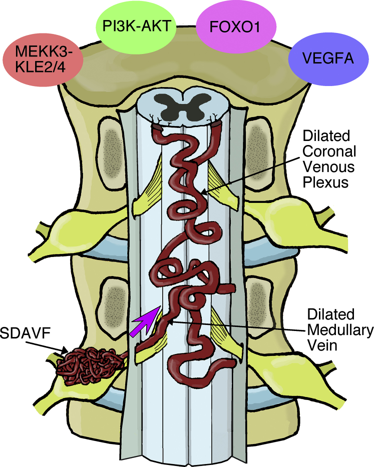
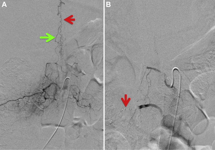
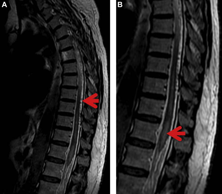
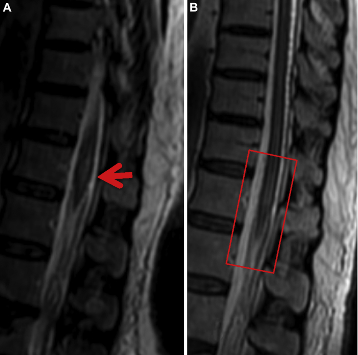
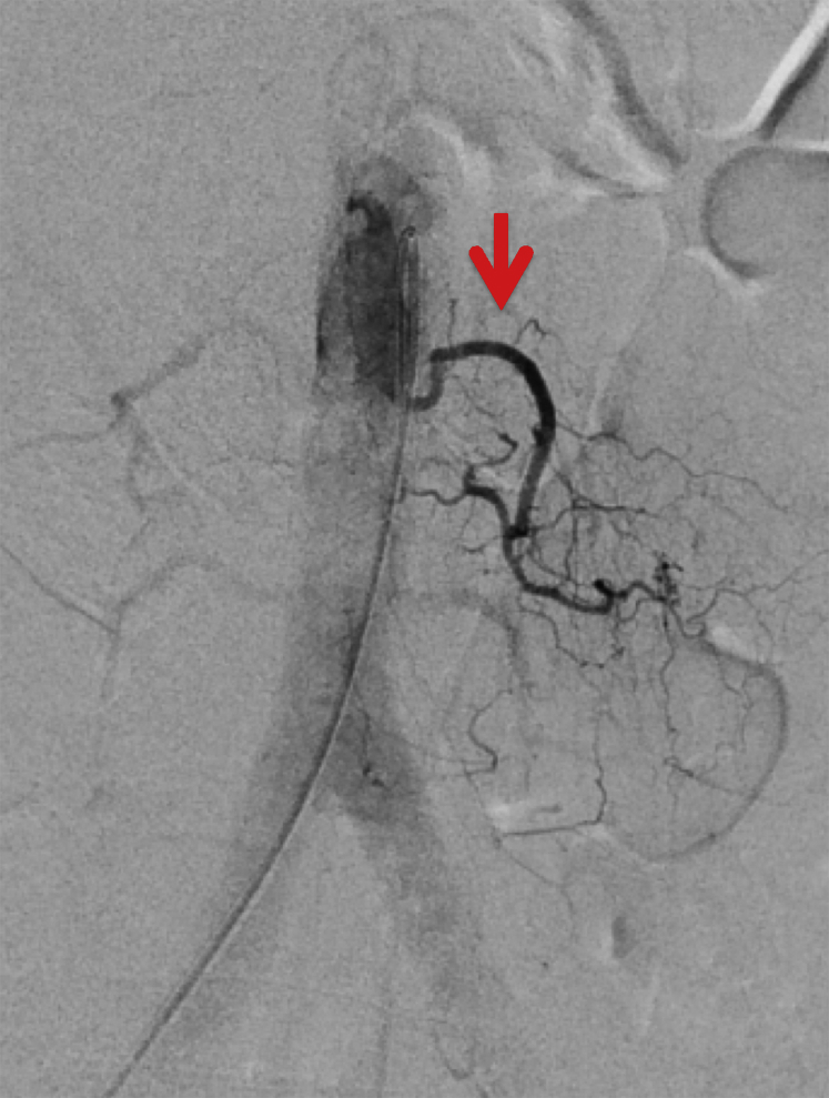
A spinal dural arteriovenous fistula is an abnormally layered connection between radicular arteries and venous plexus of the spinal cord. This vascular condition is relatively rare with an incidence of 5-10 cases per million in the general population. Diagnosis of spinal dural arteriovenous fistula is differentiated by contrast-enhanced magnetic resonance angiography or structural magnetic resonance imaging, but a definitive diagnosis requires spinal angiography methods. Here, we report a case of a 67-year-old female with a spinal dural arteriovenous fistula, provide a pertinent clinical history to the case nosology, and discuss the biology of adhesive proteins, chemotactic molecules, and transcription factors that modify the behavior of the vasculature to possibly cause sensorimotor deficits.
脊髓硬脊膜动静脉瘘是神经根动脉与脊髓静脉丛之间异常分层的连接。这种血管疾病相对罕见,在普通人群中的发病率为每百万人口5至10例。脊髓硬脊膜动静脉瘘的诊断通过对比增强磁共振血管造影或结构磁共振成像进行鉴别,但明确诊断需要采用脊髓血管造影方法。在此,我们报告一例67岁女性脊髓硬脊膜动静脉瘘病例,提供与该病例分类相关的临床病史,并讨论黏附蛋白、趋化分子和转录因子的生物学特性,这些物质可改变血管行为,可能导致感觉运动功能障碍。