Nakaoka H J, Tanei Z, Hara T, Weng J S, Kanamori A, Hayashi T, Sato H, Orimo A, Otsuji K, Tada K, Morikawa T, Sasaki T, Fukayama M, Seiki M, Murakami Y, Sakamoto T
Division of Molecular Pathology, Department of Cancer Biology, Institute of Medical Science, The University of Tokyo, Shirokanedai, Minato-ku, Tokyo, Japan.
Department of Pathology, Graduate School of Medicine, The University of Tokyo, Hongo, Bunkyo-ku, Tokyo, Japan.
Oncogenesis. 2017 May 15;6(5):e334. doi: 10.1038/oncsis.2017.27.
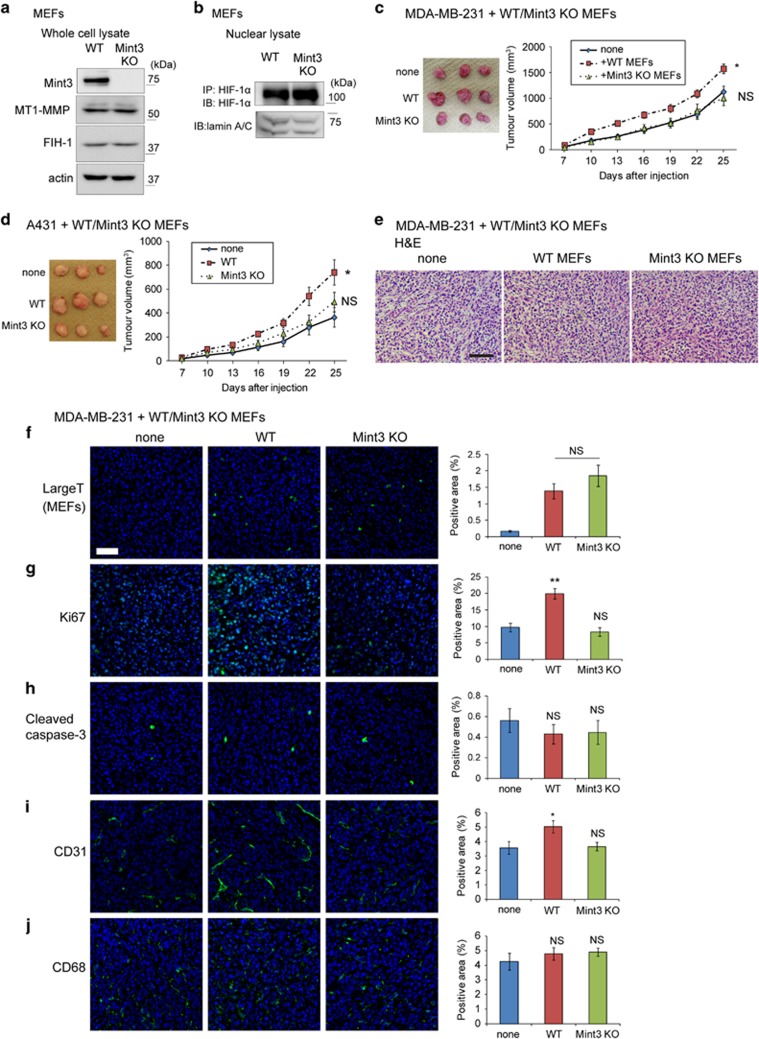
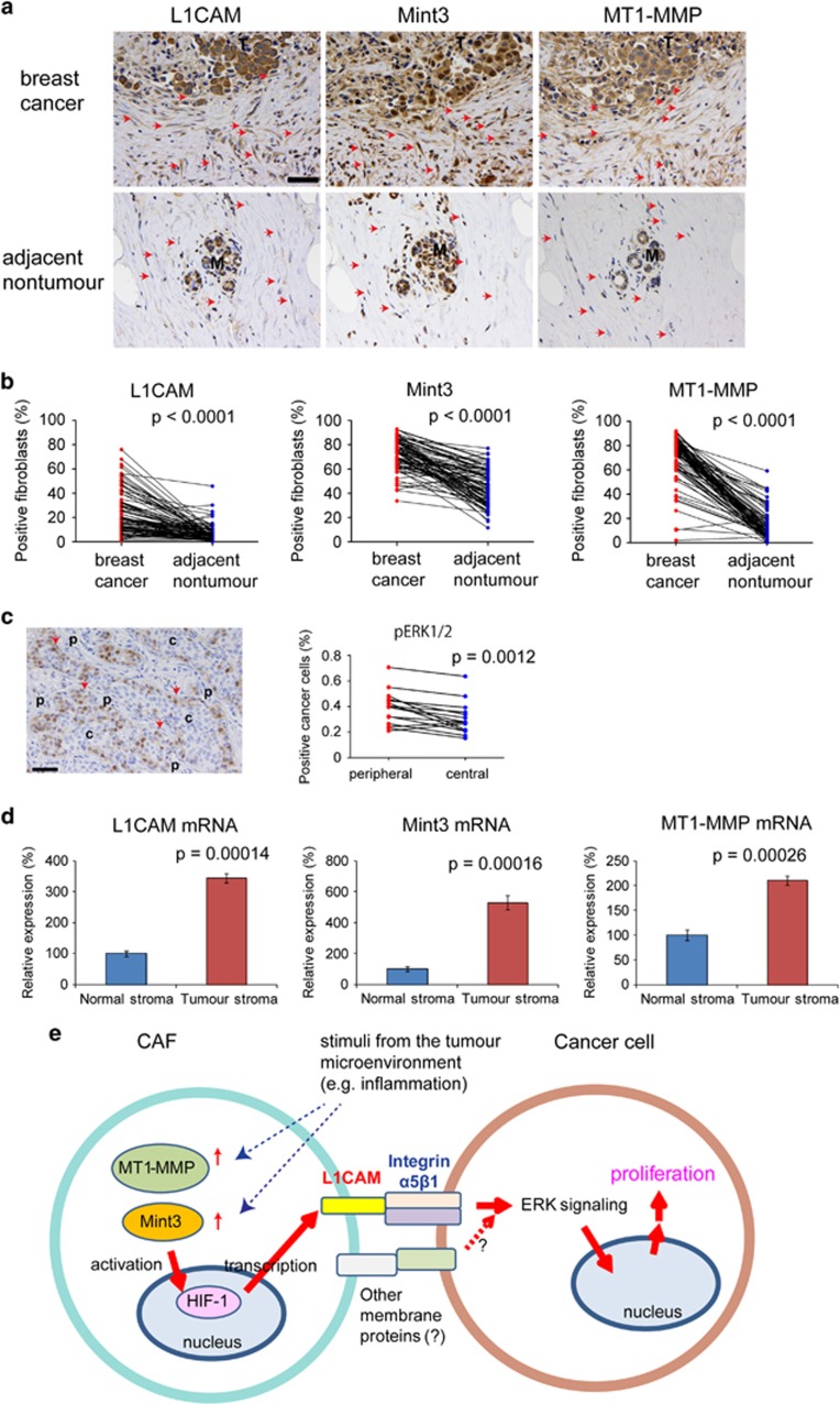
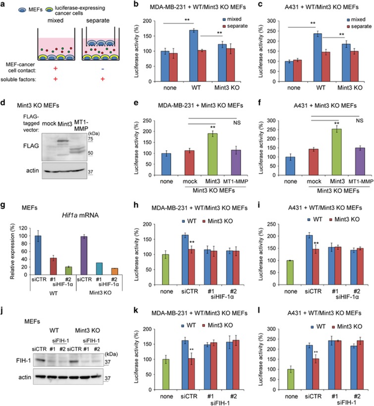
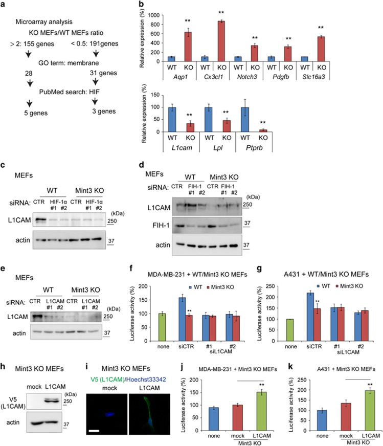
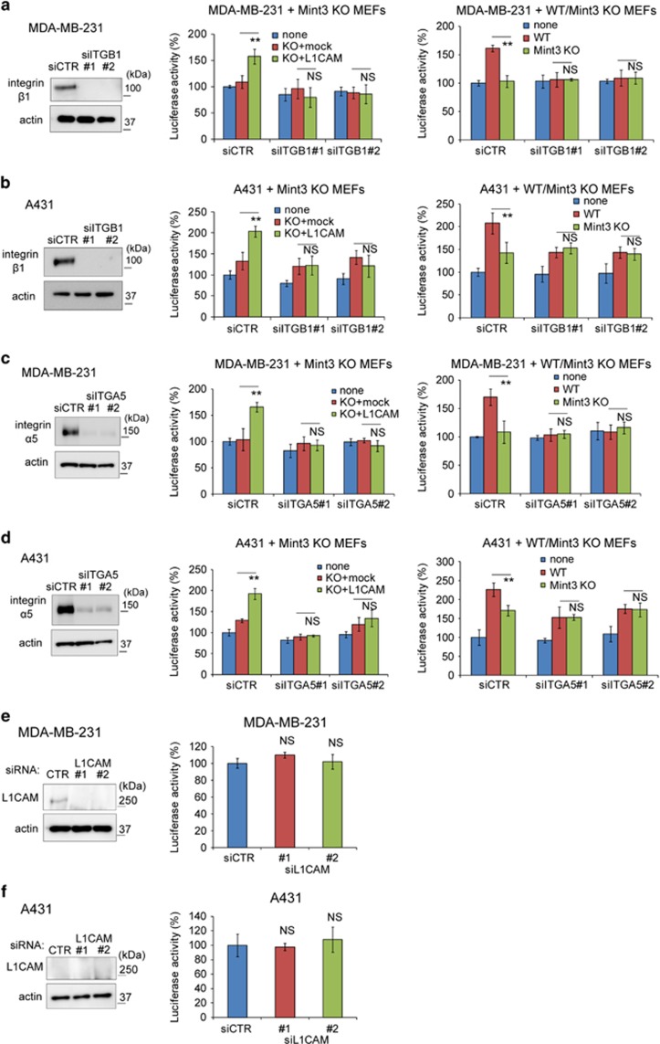
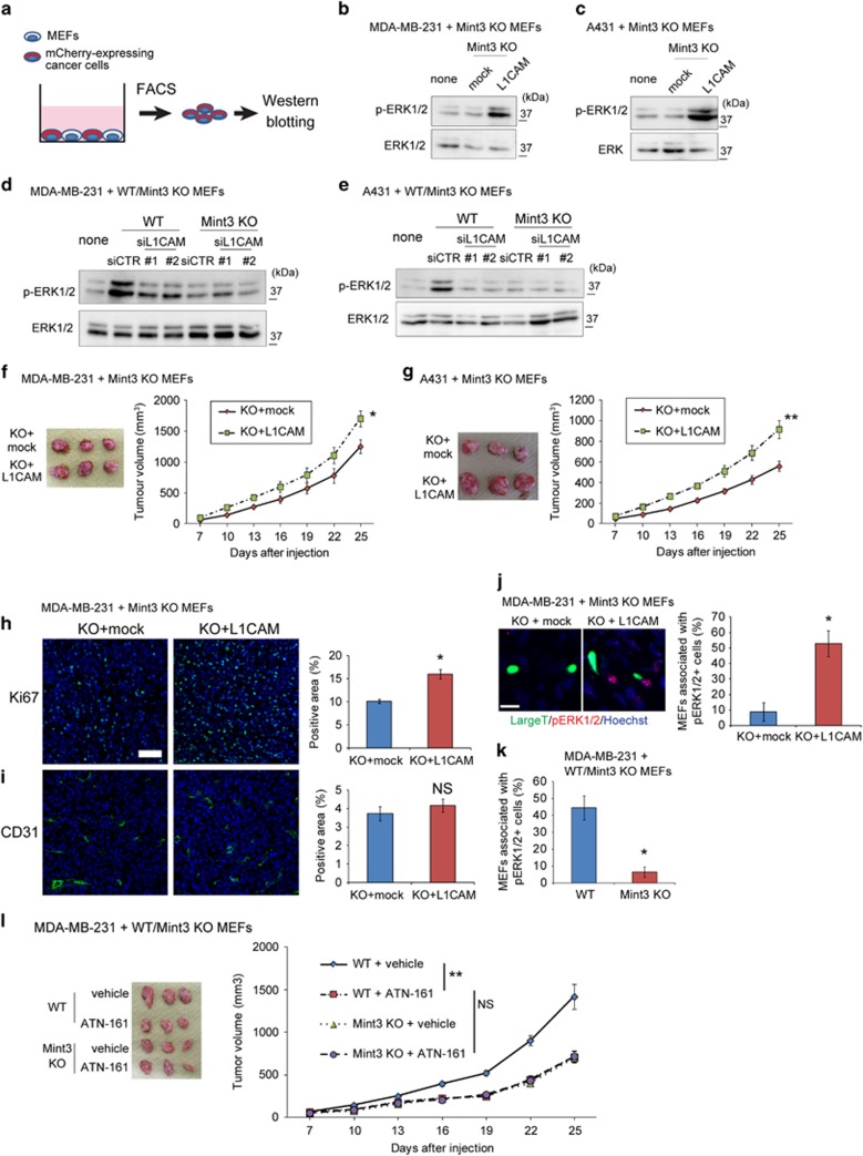
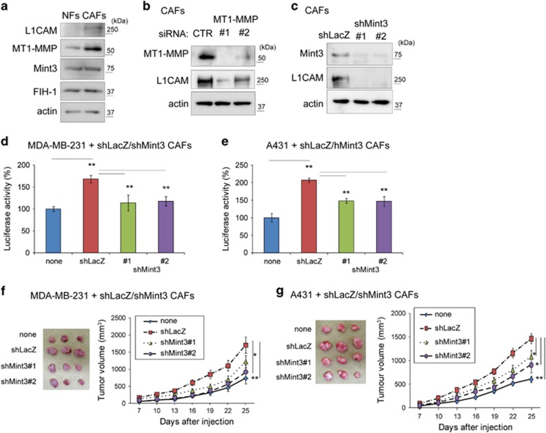
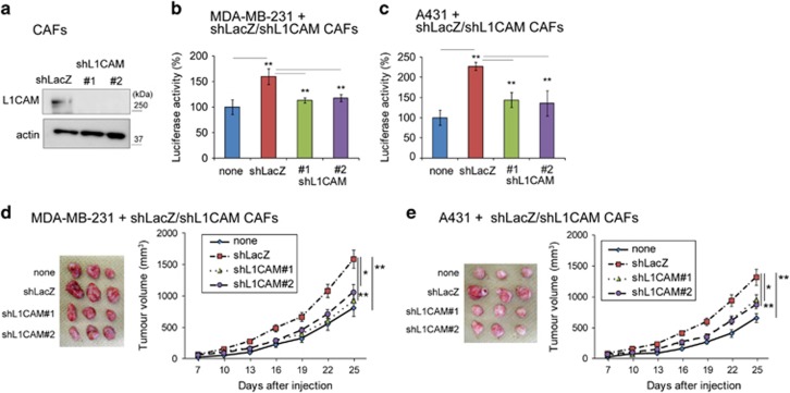
Fibroblasts are some of the major cells in tumour tissues that influence tumour progression and drug resistance. However, our understanding on fibroblast-mediated tumour malignancy remains incomplete. Munc18-1-interacting protein 3 (Mint3) is known as an activator of hypoxia-inducible factor-1 (HIF-1) even during normoxia in cancer cells, macrophages and fibroblasts. Although Mint3 promotes ATP production via glycolysis by activating HIF-1 in cancer cells and macrophages, the biological role of Mint3-mediated HIF-1 activation in fibroblasts remains unclear. To address this, we examined whether Mint3 in fibroblasts contributes to tumour growth. Mint3 depletion in mouse embryonic fibroblasts (MEFs) decreased tumour growth of co-injected human breast cancer cells, MDA-MB-231 and epidermoid carcinoma A431 cells in mice. In MEFs, Mint3 also promoted cancer cell proliferation in vitro in a cell-cell contact-dependent manner. Mint3-mediated cancer cell proliferation depended on HIF-1, and further gene expression analysis revealed that the cell adhesion molecule, L1 cell adhesion molecule (L1CAM), was induced by Mint3 and HIF-1 in fibroblasts. Mint3-mediated L1CAM expression in fibroblasts stimulated the ERK signalling pathway via integrin α5β1 in cancer cells, and promoted cancer cell proliferation in vitro and tumour growth. In cancer-associated fibroblasts (CAFs), knockdown of MT1-MMP, which promotes Mint3-mediated HIF-1 activation, or Mint3 decreased L1CAM expression. As MEFs, CAFs also promoted cancer cell proliferation in vitro, and tumour growth via Mint3 and L1CAM. In human breast cancer specimens, the number of fibroblasts expressing L1CAM, Mint3 and MT1-MMP was higher in cancer regions than in adjacent benign regions. In addition, more phospho-ERK1/2-positive cancer cells existed in the peripheral region surrounded by the stroma than in the central region of solid breast cancer nest. Thus, Mint3 in fibroblasts might be a good target for cancer therapy by regulating cancer cell-stromal cell communication.
成纤维细胞是肿瘤组织中影响肿瘤进展和耐药性的一些主要细胞。然而,我们对成纤维细胞介导的肿瘤恶性程度的理解仍不完整。Munc18-1相互作用蛋白3(Mint3)即使在癌细胞、巨噬细胞和成纤维细胞的常氧条件下也被称为缺氧诱导因子-1(HIF-1)的激活剂。尽管Mint3通过激活癌细胞和巨噬细胞中的HIF-1促进糖酵解产生ATP,但Mint3介导的HIF-1激活在成纤维细胞中的生物学作用仍不清楚。为了解决这个问题,我们研究了成纤维细胞中的Mint3是否有助于肿瘤生长。小鼠胚胎成纤维细胞(MEF)中Mint3的缺失降低了共注射的人乳腺癌细胞MDA-MB-231和表皮样癌A431细胞在小鼠体内的肿瘤生长。在MEF中,Mint3还以细胞间接触依赖的方式促进体外癌细胞增殖。Mint3介导的癌细胞增殖依赖于HIF-1,进一步的基因表达分析表明,细胞粘附分子L1细胞粘附分子(L1CAM)在成纤维细胞中由Mint3和HIF-1诱导。Mint3介导的成纤维细胞中L1CAM的表达通过癌细胞中的整合素α5β1刺激ERK信号通路,并促进体外癌细胞增殖和肿瘤生长。在癌症相关成纤维细胞(CAF)中,促进Mint3介导的HIF-1激活的MT1-MMP的敲低或Mint3降低了L1CAM的表达。与MEF一样,CAF也促进体外癌细胞增殖,并通过Mint3和L1CAM促进肿瘤生长。在人乳腺癌标本中,表达L1CAM、Mint3和MT1-MMP的成纤维细胞数量在癌区比在相邻良性区更高。此外,在实体乳腺癌巢的周边区域被基质包围的区域中,磷酸化ERK1/2阳性癌细胞比中央区域更多。因此,成纤维细胞中的Mint3可能是通过调节癌细胞-基质细胞通讯进行癌症治疗的一个良好靶点。