Adolphe Merkle Institute, University of Fribourg, Chemin des Verdiers 4, CH-1700 Fribourg, Switzerland.
Institute of Micro- and Nanostructure Research and Center for Nanoanalysis and Electron Microscopy, Department of Materials Science and Engineering, Friedrich-Alexander-Universität Erlangen-Nürnberg, Cauerstraße 6, D-91058 Erlangen, Germany.
Sci Adv. 2017 Apr 26;3(4):e1603119. doi: 10.1126/sciadv.1603119. eCollection 2017 Apr.
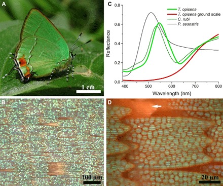
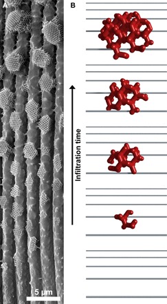
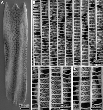
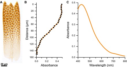
The formation of the biophotonic gyroid material in butterfly wing scales is an exceptional feat of evolutionary engineering of functional nanostructures. It is hypothesized that this nanostructure forms by chitin polymerization inside a convoluted membrane of corresponding shape in the endoplasmic reticulum. However, this dynamic formation process, including whether membrane folding and chitin expression are simultaneous or sequential processes, cannot yet be elucidated by in vivo imaging. We report an unusual hierarchical ultrastructure in the butterfly that, as a solid material, allows high-resolution three-dimensional microscopy. Rather than the conventional polycrystalline space-filling arrangement, a gyroid occurs in isolated facetted crystallites with a pronounced size gradient. When interpreted as a sequence of time-frozen snapshots of the morphogenesis, this arrangement provides insight into the formation mechanisms of the nanoporous gyroid material as well as of the intracellular organelle membrane that acts as the template.
蝴蝶翅膀鳞片中生物光子的螺旋形材料的形成是功能纳米结构进化工程的非凡壮举。据推测,这种纳米结构是通过内质网中相应形状的卷曲膜内的几丁质聚合形成的。然而,这种动态形成过程,包括膜折叠和几丁质表达是同时发生还是顺序发生的过程,目前还无法通过体内成像来阐明。我们报告了蝴蝶中一种不寻常的分层超微结构,作为一种固体材料,它允许进行高分辨率的三维显微镜观察。与传统的多晶空间填充排列不同,螺旋形出现在具有明显大小梯度的孤立面心立方结晶中。当将其解释为形态发生的时间冻结快照序列时,这种排列为纳米多孔螺旋形材料的形成机制以及作为模板的细胞内细胞器膜的形成机制提供了深入的了解。