Department of Orthopaedic Surgery, Medical University of Vienna, Waehringer Guertel 18-20, 1090, Vienna, Austria.
Department of Biomedical Imaging and Image-guided Therapy, Medical University of Vienna, Waehringer Guertel 18-20, 1090, Vienna, Austria.
Knee Surg Sports Traumatol Arthrosc. 2018 Jul;26(7):2103-2109. doi: 10.1007/s00167-017-4567-x. Epub 2017 May 15.
The aim of this study was to determine the reliability and validity of preoperative magnetic resonance imaging (MRI) scans for the detection of additional pathologies in patients with chronic ankle instability (CAI) compared to arthroscopic findings.
Preoperative MRI images of 30 patients were evaluated regarding articular and periarticular comorbidities and compared to intraoperative findings. The reliability of MRI was determined by calculating specificity, sensitivity, as well as positive and negative predictive values. The accuracy of the classification of cartilage lesions by Outerbridge and Berndt and Harty rating scales was determined by calculating the area under the receiver operating curve (AUC).
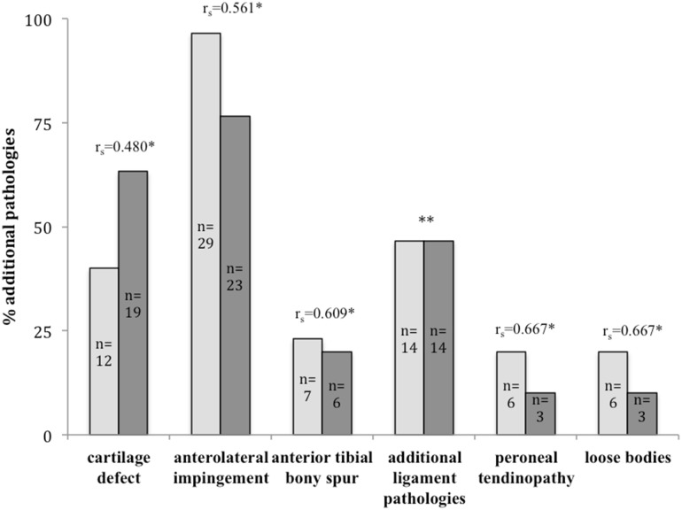
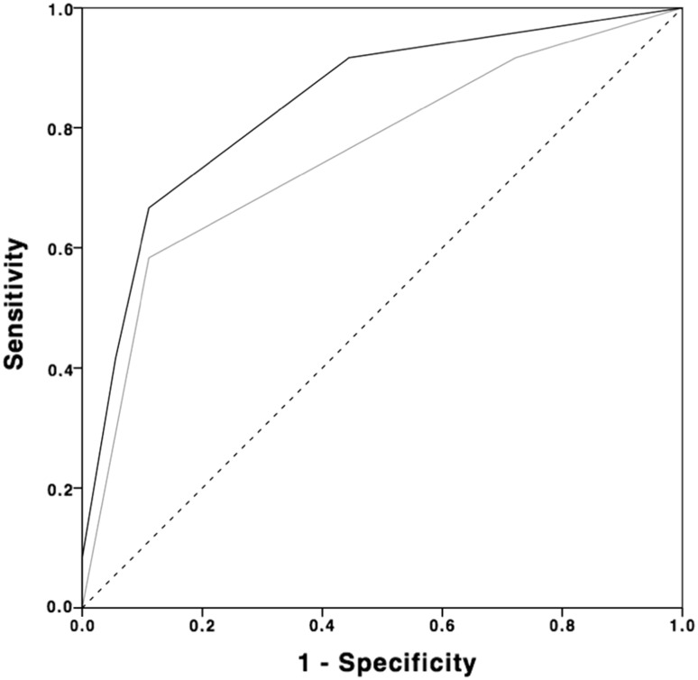
In total, 72 additional pathologies were found arthroscopically compared to 73 lesions gathered from MRI images. Sensitivity ranged from 89% for peroneal tendinopathy to 28% for additional ligamentous lesions. Specificity ranged from 100% for anterolateral impingement, loose bodies and peroneal tendinopathy to 38% for additional ligamentous lesions. For cartilage lesions, sensitivity was at 91% and specificity was at 55% for the Outerbridge grading scale. For the Berndt and Harty classification system, sensitivity was at 91% and specificity was at 28%. Correlation of additional pathologies ranged from weak (r = 0.48; p = 0.02) to moderate results (r = 0.67; p < 0.001).
CAI is associated with a high incidence of additional pathologies. In some cases, MRI delivers insufficient results, which may lead to misinterpretation of present comorbidities. MRI is a helpful tool for preoperative evaluation, but arthroscopy remains gold standard in the diagnosis of associated lesions in patients with CAI.
III.
本研究旨在比较术前磁共振成像(MRI)与关节镜检查结果,评估其在检测慢性踝关节不稳定(CAI)患者合并症方面的可靠性和有效性。
对 30 例患者的术前 MRI 图像进行评估,包括关节和关节周围的合并症,并与术中发现进行比较。通过计算特异性、敏感性、阳性和阴性预测值来确定 MRI 的可靠性。通过计算接受者操作特征曲线(AUC)下的面积来确定Outerbridge 和 Berndt 和 Harty 分级系统对软骨损伤的分类准确性。
与 MRI 图像相比,关节镜下共发现 72 种额外的病变。敏感性范围从腓骨肌腱病变的 89%到其他韧带病变的 28%。特异性范围从前外侧撞击征、游离体和腓骨肌腱病变的 100%到其他韧带病变的 38%。对于软骨病变,Outerbridge 分级的敏感性为 91%,特异性为 55%。对于 Berndt 和 Harty 分类系统,敏感性为 91%,特异性为 28%。额外病变的相关性从弱(r=0.48;p=0.02)到中度(r=0.67;p<0.001)。
CAI 常伴有较高的合并症发生率。在某些情况下,MRI 的结果不够充分,可能导致对现有合并症的误解。MRI 是术前评估的有用工具,但在诊断 CAI 患者的相关病变方面,关节镜仍然是金标准。
III。