• 文献检索
  • 文档翻译
  • 深度研究
  • 学术资讯
  • Suppr Zotero 插件Zotero 插件
  • 邀请有礼
  • 套餐&价格
  • 历史记录
应用&插件
Suppr Zotero 插件Zotero 插件浏览器插件Mac 客户端Windows 客户端微信小程序
定价
高级版会员购买积分包购买API积分包
服务
文献检索文档翻译深度研究API 文档MCP 服务
关于我们
关于 Suppr公司介绍联系我们用户协议隐私条款
关注我们

Suppr 超能文献

核心技术专利:CN118964589B侵权必究
粤ICP备2023148730 号-1Suppr @ 2026

文献检索

告别复杂PubMed语法,用中文像聊天一样搜索,搜遍4000万医学文献。AI智能推荐,让科研检索更轻松。

立即免费搜索

文件翻译

保留排版,准确专业,支持PDF/Word/PPT等文件格式,支持 12+语言互译。

免费翻译文档

深度研究

AI帮你快速写综述,25分钟生成高质量综述,智能提取关键信息,辅助科研写作。

立即免费体验

蛋白质结构研究中酪氨酸侧基的紫外-可见光谱。第2部分:选定的应用。

UV-Vis spectroscopy of tyrosine side-groups in studies of protein structure. Part 2: selected applications.

作者信息

Antosiewicz Jan M, Shugar David

机构信息

Division of Biophysics, Institute of Experimental Physics, Faculty of Physics, University of Warsaw, żwirki i Wigury 93, 02-089, Warsaw, Poland.

Institute of Biochemistry & Biophysics, Polish Academy of Sciences, Pawinskiego 5a, 02-106, Warsaw, Poland.

出版信息

Biophys Rev. 2016 Jun;8(2):163-177. doi: 10.1007/s12551-016-0197-7. Epub 2016 May 4.

DOI:10.1007/s12551-016-0197-7
PMID:28510057
原文链接:https://pmc.ncbi.nlm.nih.gov/articles/PMC4884208/
Abstract

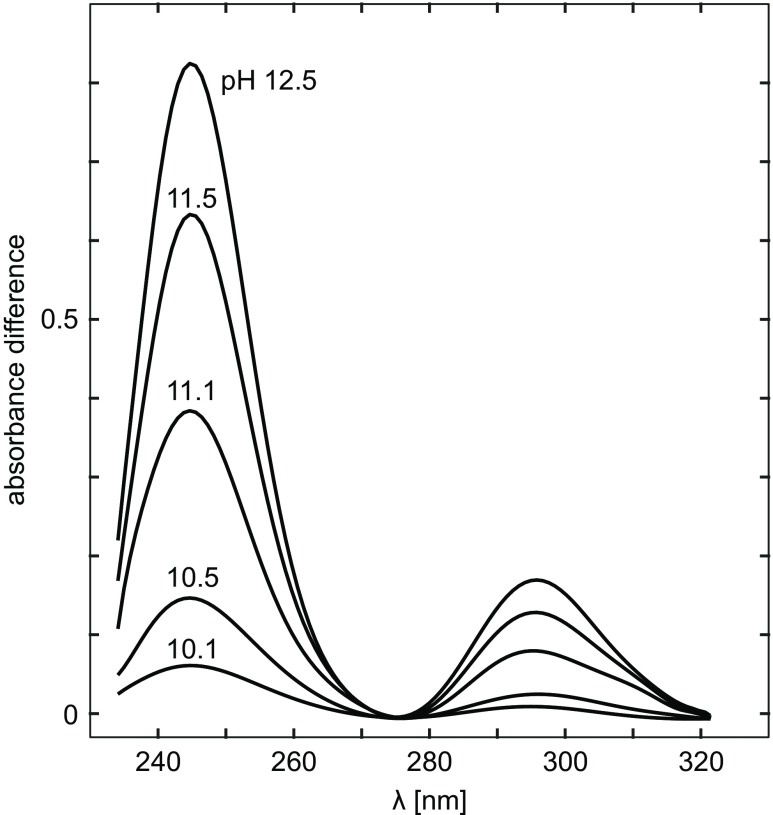
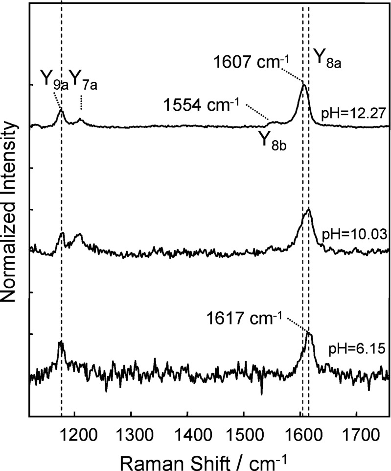
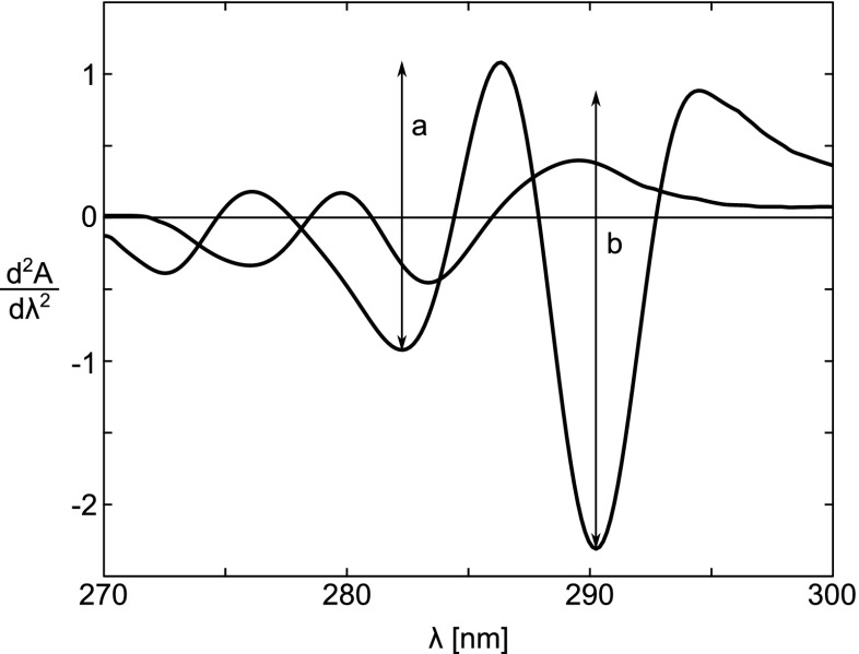
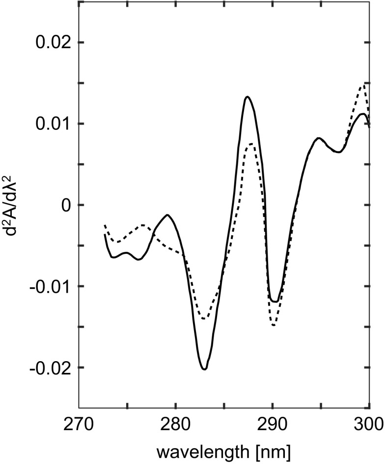
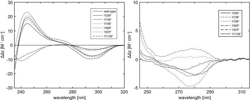
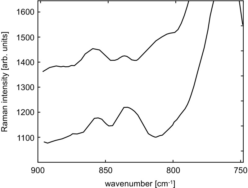
In Part 2 we discuss application of several different types of UV-Vis spectroscopy, such as normal, difference, and second-derivative UV absorption spectroscopy, fluorescence spectroscopy, linear and circular dichroism spectroscopy, and Raman spectroscopy, of the side-chain of tyrosine residues in different molecular environments. We review the ways these spectroscopies can be used to probe complex protein structures.

摘要

在第2部分中,我们讨论了几种不同类型的紫外可见光谱在不同分子环境中酪氨酸残基侧链上的应用,如常规紫外吸收光谱、差示紫外吸收光谱、二阶导数紫外吸收光谱、荧光光谱、线性和圆二色光谱以及拉曼光谱。我们回顾了这些光谱用于探测复杂蛋白质结构的方法。

https://cdn.ncbi.nlm.nih.gov/pmc/blobs/3a0c/5418476/b5c10532c881/12551_2016_197_Fig18_HTML.jpg
https://cdn.ncbi.nlm.nih.gov/pmc/blobs/3a0c/5418476/c1dcf67c9714/12551_2016_197_Fig1_HTML.jpg
https://cdn.ncbi.nlm.nih.gov/pmc/blobs/3a0c/5418476/51256f134ec4/12551_2016_197_Fig2_HTML.jpg
https://cdn.ncbi.nlm.nih.gov/pmc/blobs/3a0c/5418476/2519a9e3e702/12551_2016_197_Fig3_HTML.jpg
https://cdn.ncbi.nlm.nih.gov/pmc/blobs/3a0c/5418476/245ad4ee034f/12551_2016_197_Fig4_HTML.jpg
https://cdn.ncbi.nlm.nih.gov/pmc/blobs/3a0c/5418476/5631425befaf/12551_2016_197_Fig5_HTML.jpg
https://cdn.ncbi.nlm.nih.gov/pmc/blobs/3a0c/5418476/03062810ff3a/12551_2016_197_Fig6_HTML.jpg
https://cdn.ncbi.nlm.nih.gov/pmc/blobs/3a0c/5418476/3ec63f3a0cbb/12551_2016_197_Fig7_HTML.jpg
https://cdn.ncbi.nlm.nih.gov/pmc/blobs/3a0c/5418476/62200337c444/12551_2016_197_Fig8_HTML.jpg
https://cdn.ncbi.nlm.nih.gov/pmc/blobs/3a0c/5418476/41e5d1721566/12551_2016_197_Fig9_HTML.jpg
https://cdn.ncbi.nlm.nih.gov/pmc/blobs/3a0c/5418476/5138639a7b3d/12551_2016_197_Fig10_HTML.jpg
https://cdn.ncbi.nlm.nih.gov/pmc/blobs/3a0c/5418476/ecaf07be8309/12551_2016_197_Fig11_HTML.jpg
https://cdn.ncbi.nlm.nih.gov/pmc/blobs/3a0c/5418476/af0af8055d75/12551_2016_197_Fig12_HTML.jpg
https://cdn.ncbi.nlm.nih.gov/pmc/blobs/3a0c/5418476/0dba16ec49a6/12551_2016_197_Fig13_HTML.jpg
https://cdn.ncbi.nlm.nih.gov/pmc/blobs/3a0c/5418476/1e7a1d2f0245/12551_2016_197_Fig14_HTML.jpg
https://cdn.ncbi.nlm.nih.gov/pmc/blobs/3a0c/5418476/65ab3d87e07c/12551_2016_197_Fig15_HTML.jpg
https://cdn.ncbi.nlm.nih.gov/pmc/blobs/3a0c/5418476/9026046d4f6c/12551_2016_197_Fig16_HTML.jpg
https://cdn.ncbi.nlm.nih.gov/pmc/blobs/3a0c/5418476/df5bc29c9519/12551_2016_197_Fig17_HTML.jpg
https://cdn.ncbi.nlm.nih.gov/pmc/blobs/3a0c/5418476/b5c10532c881/12551_2016_197_Fig18_HTML.jpg

相似文献

1
UV-Vis spectroscopy of tyrosine side-groups in studies of protein structure. Part 2: selected applications.蛋白质结构研究中酪氨酸侧基的紫外-可见光谱。第2部分:选定的应用。
Biophys Rev. 2016 Jun;8(2):163-177. doi: 10.1007/s12551-016-0197-7. Epub 2016 May 4.
2
UV-Vis spectroscopy of tyrosine side-groups in studies of protein structure. Part 1: basic principles and properties of tyrosine chromophore.蛋白质结构研究中酪氨酸侧链基团的紫外可见光谱。第1部分:酪氨酸发色团的基本原理和性质。
Biophys Rev. 2016 Jun;8(2):151-161. doi: 10.1007/s12551-016-0198-6. Epub 2016 May 4.
3
UV-Vis-NIR Absorption Spectroscopy and Catalysis.紫外-可见-近红外吸收光谱与催化
Chem Rev. 2024 Mar 13;124(5):2352-2418. doi: 10.1021/acs.chemrev.3c00602. Epub 2024 Feb 26.
4
Tea types classification with data fusion of UV-Vis, synchronous fluorescence and NIR spectroscopies and chemometric analysis.利用 UV-Vis、同步荧光和近红外光谱学以及化学计量学分析的数据融合对茶叶类型进行分类。
Spectrochim Acta A Mol Biomol Spectrosc. 2019 Mar 15;211:195-202. doi: 10.1016/j.saa.2018.11.063. Epub 2018 Dec 1.
5
[Application and development of spectroscopy methodologies in the study on non-covalent interactions].[光谱学方法在非共价相互作用研究中的应用与进展]
Guang Pu Xue Yu Guang Pu Fen Xi. 2009 Jan;29(1):240-3.
6
Using resonance light scattering and UV/vis absorption spectroscopy to study the interaction between gliclazide and bovine serum albumin.利用共振光散射和紫外/可见吸收光谱法研究格列齐特与牛血清白蛋白之间的相互作用。
Luminescence. 2016 Aug;31(5):1109-14. doi: 10.1002/bio.3079. Epub 2015 Dec 14.
7
UV/Vis photochemistry database: Structure, content and applications.紫外/可见光谱光化学数据库:结构、内容及应用
J Quant Spectrosc Radiat Transf. 2020 Sep 1;253. doi: 10.1016/j.jqsrt.2020.107056.
8
Comparative Study of the Interactions between Ovalbumin and five Antioxidants by Spectroscopic Methods.比较研究卵清蛋白与五种抗氧化剂的相互作用的光谱方法。
J Fluoresc. 2017 Jan;27(1):213-225. doi: 10.1007/s10895-016-1948-3. Epub 2016 Oct 8.
9
SPECTRAL METHODS FOR STUDY OF THE G-PROTEIN-COUPLED RECEPTOR RHODOPSIN. I. VIBRATIONAL AND ELECTRONIC SPECTROSCOPY.用于研究G蛋白偶联受体视紫红质的光谱方法。I. 振动光谱与电子光谱
Opt Spectrosc. 2015 May;118(5):711-717. doi: 10.1134/S0030400X15050240. Epub 2015 May 27.
10
Online Monitoring of Solutions Within Microfluidic Chips: Simultaneous Raman and UV-Vis Absorption Spectroscopies.微流控芯片内溶液的在线监测:同时拉曼和紫外-可见吸收光谱法。
ACS Sens. 2019 Sep 27;4(9):2288-2295. doi: 10.1021/acssensors.9b00736. Epub 2019 Sep 6.

引用本文的文献

1
Dual-Oriented Targeted Nanostructured SERS Label-Free Immunosensor for Detection, Quantification, and Analysis of Breast Cancer Biomarker Concentrations in Blood Serum.用于检测、定量和分析血清中乳腺癌生物标志物浓度的双导向靶向纳米结构表面增强拉曼散射无标记免疫传感器。
Biosensors (Basel). 2025 Jul 11;15(7):447. doi: 10.3390/bios15070447.
2
Utilization of Industrial Hemp Biomass Waste (I): Stability of Cannabidiol in Pre and Post- Encapsulation States.工业大麻生物质废料的利用(I):大麻二酚在包封前后状态下的稳定性
Molecules. 2025 May 10;30(10):2116. doi: 10.3390/molecules30102116.
3
Sustainable streptomycin-based Ag nanoparticle synthesis and study of the cytotoxicity.

本文引用的文献

1
Transthyretin: a multifaceted protein.转甲状腺素蛋白:一种多面蛋白。
Biomol Concepts. 2014 Mar;5(1):45-54. doi: 10.1515/bmc-2013-0038.
2
pH-dependent random coil (1)H, (13)C, and (15)N chemical shifts of the ionizable amino acids: a guide for protein pK a measurements.可电离氨基酸的pH依赖性无规卷曲(1)H、(13)C和(15)N化学位移:蛋白质pKa测量指南
J Biomol NMR. 2014 Nov;60(2-3):109-29. doi: 10.1007/s10858-014-9862-y. Epub 2014 Sep 20.
3
UV transition moments of tyrosine.酪氨酸的紫外跃迁矩。
基于链霉素的可持续银纳米颗粒合成及其细胞毒性研究。
Naunyn Schmiedebergs Arch Pharmacol. 2025 Feb 5. doi: 10.1007/s00210-025-03859-9.
4
Exploring the effect of natural deep eutectic solvents on zein: Structural and functional properties.探索天然低共熔溶剂对玉米醇溶蛋白的影响:结构和功能特性
Curr Res Food Sci. 2024 Dec 20;10:100965. doi: 10.1016/j.crfs.2024.100965. eCollection 2025.
5
Antiplasmodial potential of compounds isolated from Ziziphus mucronata and their binding to Plasmodium falciparum HGXPRT using biophysical and molecular docking studies.从枣(Ziziphus mucronata)中分离出的化合物的抗疟潜力及其通过生物物理和分子对接研究与恶性疟原虫HGXPRT的结合
Naunyn Schmiedebergs Arch Pharmacol. 2025 May;398(5):5453-5463. doi: 10.1007/s00210-024-03611-9. Epub 2024 Nov 19.
6
Valorization of treated wastewater from the soaking of baby alpaca skin fur.羊驼幼崽皮草浸泡处理后废水的资源化利用
Heliyon. 2024 Sep 21;10(18):e38247. doi: 10.1016/j.heliyon.2024.e38247. eCollection 2024 Sep 30.
7
Detection of Phenylalanine by Iridium Nanoclusters Using Time-Dependent Density Functional Theory Calculations.利用含时密度泛函理论计算通过铱纳米团簇检测苯丙氨酸
ACS Omega. 2024 Aug 28;9(36):38186-38194. doi: 10.1021/acsomega.4c05684. eCollection 2024 Sep 10.
8
Targeted FT-NIR and SERS Detection of Breast Cancer HER-II Biomarkers in Blood Serum Using PCB-Based Plasmonic Active Nanostructured Thin Film Label-Free Immunosensor Immobilized with Directional GNU-Conjugated Antibody.基于 PCB 的等离子体活性纳米结构薄膜标记自由免疫传感器,采用定向 GNU 共轭抗体固定,用于血清中乳腺癌 HER-II 生物标志物的靶向 FT-NIR 和 SERS 检测。
Sensors (Basel). 2024 Aug 20;24(16):5378. doi: 10.3390/s24165378.
9
Protopine and Allocryptopine Interactions with Plasma Proteins.原小檗碱和阿朴可托品与血浆蛋白的相互作用。
Int J Mol Sci. 2024 May 15;25(10):5398. doi: 10.3390/ijms25105398.
10
Optimized Ultraviolet-C Processing Inactivates Pathogenic and Spoilage-Associated Bacteria while Preserving Bioactive Proteins, Vitamins, and Lipids in Human Milk.优化的紫外线-C 处理在灭活致病性和腐败相关细菌的同时,保留人乳中的生物活性蛋白质、维生素和脂质。
J Agric Food Chem. 2024 May 29;72(21):12198-12208. doi: 10.1021/acs.jafc.4c02120. Epub 2024 May 16.
J Phys Chem B. 2014 Aug 7;118(31):9247-57. doi: 10.1021/jp5065352. Epub 2014 Jul 29.
4
Understanding and applying tyrosine biochemical diversity.理解并应用酪氨酸的生化多样性。
Mol Biosyst. 2014 May;10(5):952-69. doi: 10.1039/c4mb00018h.
5
Insulin: a small protein with a long journey.胰岛素:一种具有漫长旅程的小蛋白质。
Protein Cell. 2010 Jun;1(6):537-51. doi: 10.1007/s13238-010-0069-z.
6
Tyrosine autofluorescence as a measure of bovine insulin fibrillation.酪氨酸自发荧光作为牛胰岛素纤维化的测量指标。
Biophys J. 2009 Nov 4;97(9):2521-31. doi: 10.1016/j.bpj.2009.07.064.
7
A new look at the human Rad51 protein.对人类Rad51蛋白的新认识。
Proc Natl Acad Sci U S A. 2009 Aug 11;106(32):13147-8. doi: 10.1073/pnas.0906816106. Epub 2009 Aug 5.
8
Structure of human Rad51 protein filament from molecular modeling and site-specific linear dichroism spectroscopy.基于分子建模和位点特异性线性二色光谱法的人源Rad51蛋白丝结构
Proc Natl Acad Sci U S A. 2009 Aug 11;106(32):13248-53. doi: 10.1073/pnas.0902723106. Epub 2009 Jul 8.
9
UV resonance Raman spectroscopy of TTR(105-115): determination of the pKa of tyrosine.转甲状腺素蛋白(105 - 115)的紫外共振拉曼光谱:酪氨酸pKa的测定
Phys Chem Chem Phys. 2009 Jul 14;11(26):5302-9. doi: 10.1039/b823363b. Epub 2009 Apr 23.
10
Probing protein structure and dynamics by second-derivative ultraviolet absorption analysis of cation-{pi} interactions.通过阳离子-π相互作用的二阶紫外吸收分析探测蛋白质结构和动力学
Protein Sci. 2006 Oct;15(10):2228-43. doi: 10.1110/ps.062133706. Epub 2006 Sep 8.