• 文献检索
  • 文档翻译
  • 深度研究
  • 学术资讯
  • Suppr Zotero 插件Zotero 插件
  • 邀请有礼
  • 套餐&价格
  • 历史记录
应用&插件
Suppr Zotero 插件Zotero 插件浏览器插件Mac 客户端Windows 客户端微信小程序
定价
高级版会员购买积分包购买API积分包
服务
文献检索文档翻译深度研究API 文档MCP 服务
关于我们
关于 Suppr公司介绍联系我们用户协议隐私条款
关注我们

Suppr 超能文献

核心技术专利:CN118964589B侵权必究
粤ICP备2023148730 号-1Suppr @ 2026

文献检索

告别复杂PubMed语法,用中文像聊天一样搜索,搜遍4000万医学文献。AI智能推荐,让科研检索更轻松。

立即免费搜索

文件翻译

保留排版,准确专业,支持PDF/Word/PPT等文件格式,支持 12+语言互译。

免费翻译文档

深度研究

AI帮你快速写综述,25分钟生成高质量综述,智能提取关键信息,辅助科研写作。

立即免费体验

基于光学相干断层扫描血管造影术测量人体皮肤血管密度的可重复性

Repeatability of vessel density measurement in human skin by OCT-based microangiography.

作者信息

Men S J, Chen C-L, Wei W, Lai T-Y, Song S Z, Wang R K

机构信息

Department of Bioengineering, University of Washington, Seattle, WA, USA.

出版信息

Skin Res Technol. 2017 Nov;23(4):607-612. doi: 10.1111/srt.12379. Epub 2017 May 17.

DOI:10.1111/srt.12379
PMID:28514014
原文链接:https://pmc.ncbi.nlm.nih.gov/articles/PMC5632107/
Abstract

PURPOSE

To investigate the repeatability of vessel density measurement at human arm skin in healthy subjects with OCT-based microangiography (OMAG).

METHODS

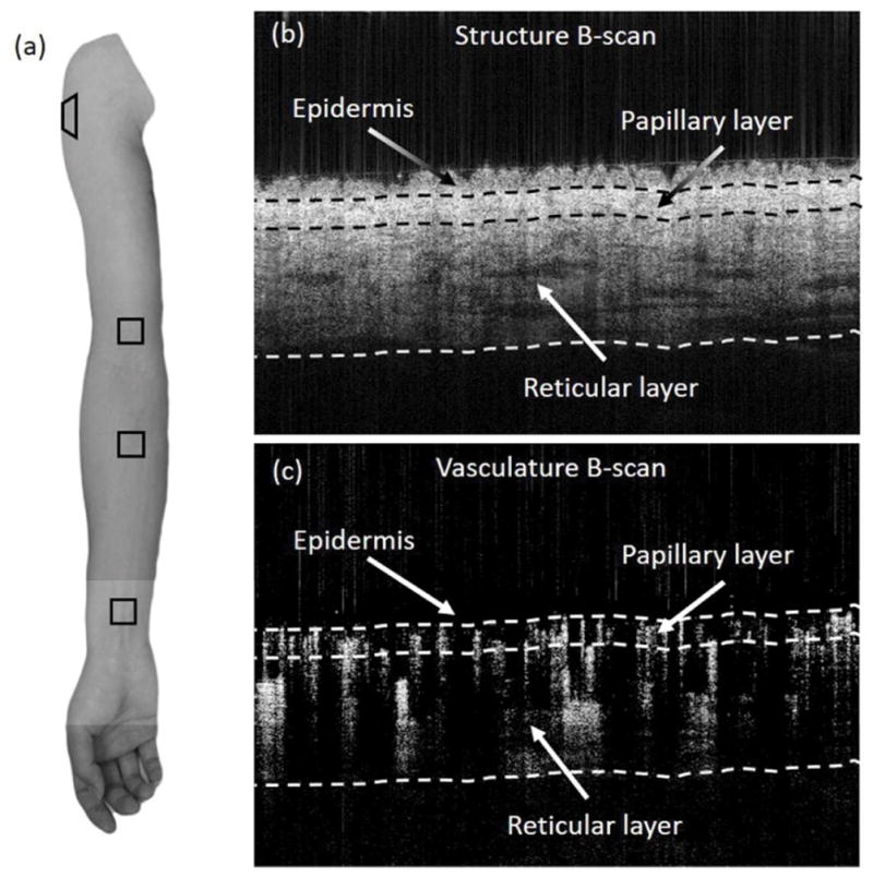
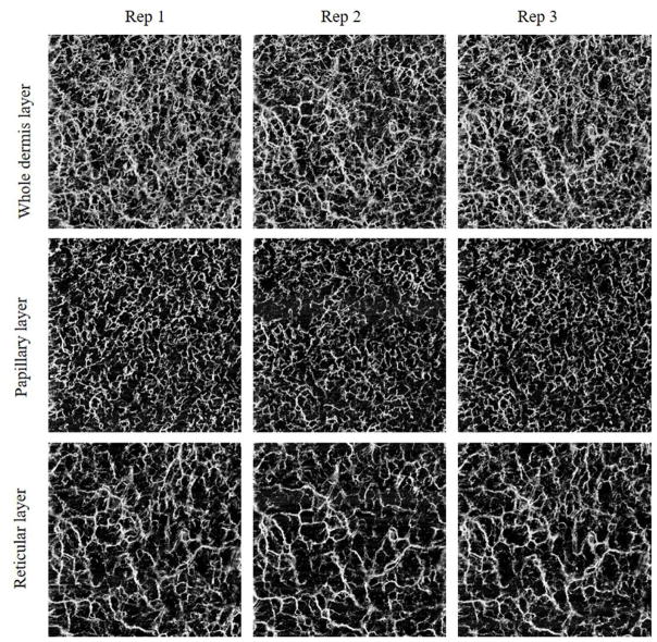
Four locations including volar wrist, volar forearm, shoulder, and volar upper arm were scanned using an optimized swept source OCT system, working at center wavelength of 1300 nm and A-line rate of 100 kHz. Three scans were acquired at each location at the same visit. Vascular images of papillary dermis, reticular dermis, and the whole dermis layer were generated with OMAG processing and automatic segmentation algorithms. The vessel density (VD) of each layer was calculated based on vascular images, and the repeatability of the VD at the same physiological location was thereafter assessed.

RESULTS

Fifteen healthy volunteers were included. High repeatability of VD was found for wrist, forearm, shoulder, and upper arm (coefficient of variation (CV)=2.4, 2.7, 2.7, 2.0, and intraclass correlation coefficient (ICC)=0.906, 0.854, 0.943, 0.916 respectively). The VD measurements showed no significant difference between the four locations in any of the three layers, ie papillary layer (P=.1063), reticular layer (P=.3371), and whole dermis layer (P=.3233).

CONCLUSION

Quantification of VD by using OCT/OMAG is repeatable when imaging skin tissue beds in healthy individuals.

摘要

目的

利用基于光学相干断层扫描的微血管造影术(OMAG)研究健康受试者手臂皮肤血管密度测量的可重复性。

方法

使用优化的扫频源光学相干断层扫描系统对包括掌侧腕部、掌侧前臂、肩部和掌侧上臂在内的四个部位进行扫描,该系统的中心波长为1300 nm,A线速率为100 kHz。在同一次就诊时,对每个部位进行三次扫描。通过OMAG处理和自动分割算法生成乳头真皮、网状真皮和整个真皮层的血管图像。根据血管图像计算各层的血管密度(VD),然后评估同一生理部位VD的可重复性。

结果

纳入15名健康志愿者。发现腕部、前臂、肩部和上臂的VD具有高重复性(变异系数(CV)分别为2.4、2.7、2.7、2.0,组内相关系数(ICC)分别为0.906、0.854、0.943、0.916)。在乳头层(P = 0.1063)、网状层(P = 0.3371)和整个真皮层(P = 0.3233)这三层中的任何一层,四个部位之间的VD测量均无显著差异。

结论

在对健康个体的皮肤组织床进行成像时,使用OCT/OMAG对VD进行定量是可重复的。

https://cdn.ncbi.nlm.nih.gov/pmc/blobs/ce2c/5632107/32bf93223e37/nihms870743f2.jpg
https://cdn.ncbi.nlm.nih.gov/pmc/blobs/ce2c/5632107/918e0dcbc808/nihms870743f1.jpg
https://cdn.ncbi.nlm.nih.gov/pmc/blobs/ce2c/5632107/32bf93223e37/nihms870743f2.jpg
https://cdn.ncbi.nlm.nih.gov/pmc/blobs/ce2c/5632107/918e0dcbc808/nihms870743f1.jpg
https://cdn.ncbi.nlm.nih.gov/pmc/blobs/ce2c/5632107/32bf93223e37/nihms870743f2.jpg

相似文献

1
Repeatability of vessel density measurement in human skin by OCT-based microangiography.基于光学相干断层扫描血管造影术测量人体皮肤血管密度的可重复性
Skin Res Technol. 2017 Nov;23(4):607-612. doi: 10.1111/srt.12379. Epub 2017 May 17.
2
Repeatability and Reproducibility of Quantitative Assessment of the Retinal Microvasculature Using Optical Coherence Tomography Angiography Based on Optical Microangiography.基于光微脉管成像的光学相干断层扫描血管造影定量评估视网膜微血管的可重复性和再现性。
Biomed Environ Sci. 2018 Jun;31(6):407-412. doi: 10.3967/bes2018.054.
3
Repeatability and reproducibility of optic nerve head perfusion measurements using optical coherence tomography angiography.采用光学相干断层扫描血管造影术测量视盘血流灌注的可重复性和再现性。
J Biomed Opt. 2016 Jun 1;21(6):65002. doi: 10.1117/1.JBO.21.6.065002.
4
Quantifying Retinal Microvascular Changes in Uveitis Using Spectral-Domain Optical Coherence Tomography Angiography.使用频域光学相干断层扫描血管造影术量化葡萄膜炎中的视网膜微血管变化。
Am J Ophthalmol. 2016 Nov;171:101-112. doi: 10.1016/j.ajo.2016.08.035. Epub 2016 Sep 2.
5
Repeatability of automated vessel density measurements using optical coherence tomography angiography.使用光学相干断层扫描血管造影术进行自动血管密度测量的可重复性
Br J Ophthalmol. 2017 Apr;101(4):449-452. doi: 10.1136/bjophthalmol-2016-308764. Epub 2016 Jul 22.
6
High resolution imaging of acne lesion development and scarring in human facial skin using OCT-based microangiography.使用基于光学相干断层扫描(OCT)的微血管造影术对人类面部皮肤痤疮病变发展和瘢痕形成进行高分辨率成像。
Lasers Surg Med. 2015 Mar;47(3):231-8. doi: 10.1002/lsm.22339. Epub 2015 Mar 5.
7
Intrasession and Between-Visit Variability of Retinal Vessel Density Values Measured with OCT Angiography in Diabetic Patients.糖尿病患者的 OCT 血管造影测量的视网膜血管密度值的日内和访间变异性。
Sci Rep. 2018 Jul 13;8(1):10598. doi: 10.1038/s41598-018-28994-7.
8
Potential use of OCT-based microangiography in clinical dermatology.基于光学相干断层扫描的微血管造影在临床皮肤科的潜在应用。
Skin Res Technol. 2016 May;22(2):238-246. doi: 10.1111/srt.12255. Epub 2015 Sep 3.
9
Normative Data for Vascular Density in Superficial and Deep Capillary Plexuses of Healthy Adults Assessed by Optical Coherence Tomography Angiography.通过光学相干断层扫描血管造影评估健康成年人浅表和深部毛细血管丛血管密度的标准数据。
Invest Ophthalmol Vis Sci. 2016 Jul 1;57(9):OCT211-23. doi: 10.1167/iovs.15-18793.
10
A novel image processing workflow for the in vivo quantification of skin microvasculature using dynamic optical coherence tomography.一种使用动态光学相干断层扫描对皮肤微血管进行体内定量分析的新型图像处理工作流程。
Skin Res Technol. 2018 Aug;24(3):396-406. doi: 10.1111/srt.12442. Epub 2018 Feb 4.

引用本文的文献

1
Quantitative assessment of the oral microvasculature using optical coherence tomography angiography.使用光学相干断层扫描血管造影术对口腔微血管进行定量评估。
Front Bioeng Biotechnol. 2024 Sep 20;12:1464562. doi: 10.3389/fbioe.2024.1464562. eCollection 2024.
2
Pilot study of optical coherence tomography angiography-derived microvascular metrics in hands and feet of healthy and diabetic people.健康人群和糖尿病患者手部和足部的光相干断层扫描血管造影衍生微血管测量的初步研究。
Sci Rep. 2023 Jan 20;13(1):1122. doi: 10.1038/s41598-022-26871-y.
3
Development of a Vascularized Human Skin Equivalent with Hypodermis for Photoaging Studies.

本文引用的文献

1
Optical coherence tomography based angiography [Invited].基于光学相干断层扫描的血管造影术[特邀报告]
Biomed Opt Express. 2017 Jan 24;8(2):1056-1082. doi: 10.1364/BOE.8.001056. eCollection 2017 Feb 1.
2
Wide field and highly sensitive angiography based on optical coherence tomography with akinetic swept source.基于光学相干断层扫描和无运动扫频光源的宽视野高灵敏度血管造影术。
Biomed Opt Express. 2016 Dec 22;8(1):420-435. doi: 10.1364/BOE.8.000420. eCollection 2017 Jan 1.
3
Highly efficient eigen decomposition based statistical optical microangiography.
构建具有真皮下血管丛的人皮肤等效物用于光老化研究。
Biomolecules. 2022 Dec 7;12(12):1828. doi: 10.3390/biom12121828.
4
High resolution imaging and quantification of the nailfold microvasculature using optical coherence tomography angiography (OCTA) and capillaroscopy: a preliminary study in healthy subjects.使用光学相干断层扫描血管造影(OCTA)和毛细血管镜对甲襞微血管进行高分辨率成像和定量分析:一项在健康受试者中的初步研究。
Quant Imaging Med Surg. 2022 Mar;12(3):1844-1858. doi: 10.21037/qims-21-672.
5
OCTAVA: An open-source toolbox for quantitative analysis of optical coherence tomography angiography images.OCTAVA:用于光学相干断层扫描血管造影图像定量分析的开源工具箱。
PLoS One. 2021 Dec 9;16(12):e0261052. doi: 10.1371/journal.pone.0261052. eCollection 2021.
6
Imaging Motion: A Comprehensive Review of Optical Coherence Tomography Angiography.《影像中的动态:光学相干断层扫描血管成像技术全面综述》。
Adv Exp Med Biol. 2021;1310:343-365. doi: 10.1007/978-981-33-6064-8_12.
7
Automatic 3D adaptive vessel segmentation based on linear relationship between intensity and complex-decorrelation in optical coherence tomography angiography.基于光学相干断层扫描血管造影中强度与复相关之间线性关系的自动三维自适应血管分割
Quant Imaging Med Surg. 2021 Mar;11(3):895-906. doi: 10.21037/qims-20-868.
8
Polarization sensitive optical coherence tomography for imaging microvascular information within living tissue without polarization-induced artifacts.用于在无偏振诱导伪影的情况下对活体组织内微血管信息进行成像的偏振敏感光学相干断层扫描技术。
Biomed Opt Express. 2020 Oct 15;11(11):6379-6388. doi: 10.1364/BOE.403933. eCollection 2020 Nov 1.
9
Automatic skin lesion area determination of basal cell carcinoma using optical coherence tomography angiography and a skeletonization approach: Preliminary results.基于光学相干断层扫描血管造影和骨架化方法的基底细胞癌自动皮损面积测定:初步结果。
J Biophotonics. 2019 Sep;12(9):e201900131. doi: 10.1002/jbio.201900131. Epub 2019 Jun 18.
10
Toward quantitative and reproducible clinical use of OCT-Angiography.朝向 OCT 血管造影定量且可重现的临床应用。
PLoS One. 2018 Jul 6;13(7):e0197588. doi: 10.1371/journal.pone.0197588. eCollection 2018.
基于高效特征分解的统计光学微血管造影术
Quant Imaging Med Surg. 2016 Oct;6(5):557-563. doi: 10.21037/qims.2016.10.03.
4
Automated segmentation and enhancement of optical coherence tomography-acquired images of rodent brain.啮齿动物大脑光学相干断层扫描获取图像的自动分割与增强
J Neurosci Methods. 2016 Sep 1;270:132-137. doi: 10.1016/j.jneumeth.2016.06.014. Epub 2016 Jun 17.
5
Quantitative assessment of the retinal microvasculature using optical coherence tomography angiography.利用光相干断层扫描血管造影术对视网膜微血管进行定量评估。
J Biomed Opt. 2016 Jun 1;21(6):66008. doi: 10.1117/1.JBO.21.6.066008.
6
In vivo photoacoustic microscopy of human cuticle microvasculature with single-cell resolution.利用单细胞分辨率对人体角质层微血管进行体内光声显微镜成像。
J Biomed Opt. 2016 May 1;21(5):56004. doi: 10.1117/1.JBO.21.5.056004.
7
OCT-based label-free in vivo lymphangiography within human skin and areola.基于光学相干断层扫描的人体皮肤和乳晕内无标记体内淋巴管造影。
Sci Rep. 2016 Feb 19;6:21122. doi: 10.1038/srep21122.
8
An EULAR study group pilot study on reliability of simple capillaroscopic definitions to describe capillary morphology in rheumatic diseases.欧洲抗风湿病联盟(EULAR)研究小组关于描述风湿病毛细血管形态的简单毛细血管镜定义可靠性的初步研究。
Rheumatology (Oxford). 2016 May;55(5):883-90. doi: 10.1093/rheumatology/kev441. Epub 2016 Feb 3.
9
Methods and algorithms for optical coherence tomography-based angiography: a review and comparison.基于光学相干断层扫描血管造影的方法与算法:综述与比较
J Biomed Opt. 2015 Oct;20(10):100901. doi: 10.1117/1.JBO.20.10.100901.
10
Potential use of OCT-based microangiography in clinical dermatology.基于光学相干断层扫描的微血管造影在临床皮肤科的潜在应用。
Skin Res Technol. 2016 May;22(2):238-246. doi: 10.1111/srt.12255. Epub 2015 Sep 3.