Chen Xianzhuo, Wu Yi, Tao Ling, Yan Yan, Pang Jun, Zhang Shaoxiang, Li Shirong
Department of Plastic and Reconstructive Surgery, Southwest Hospital, Third Military Medical University, Chongqing, China (mainland).
Institute of Digital Medicine, College of Biomedical Engineering, Third Military Medical University, Chongqing, China (mainland).
Med Sci Monit. 2017 May 22;23:2436-2444. doi: 10.12659/msm.901926.
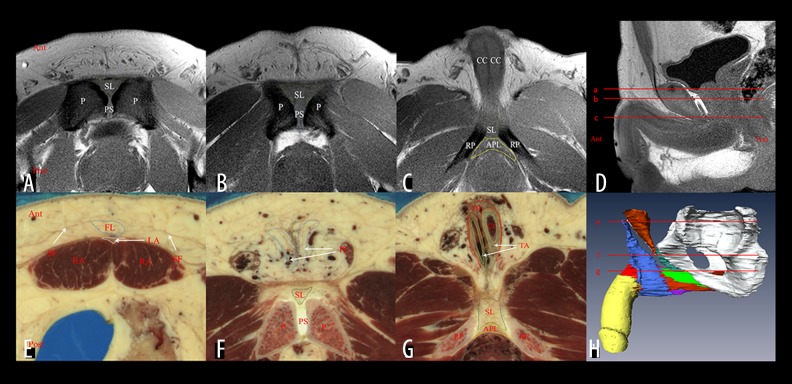
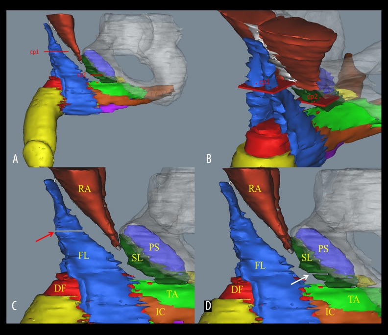
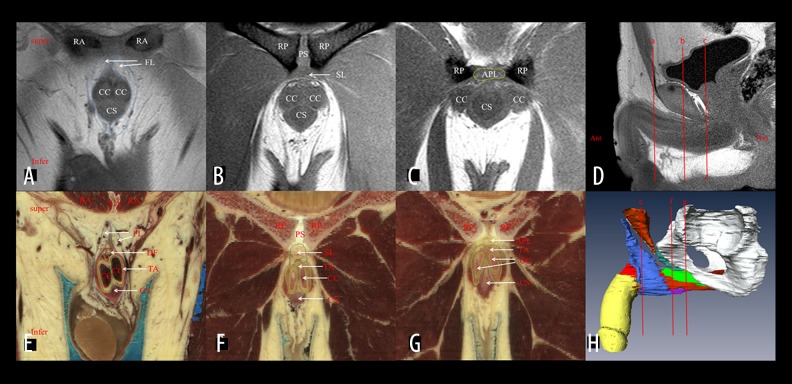
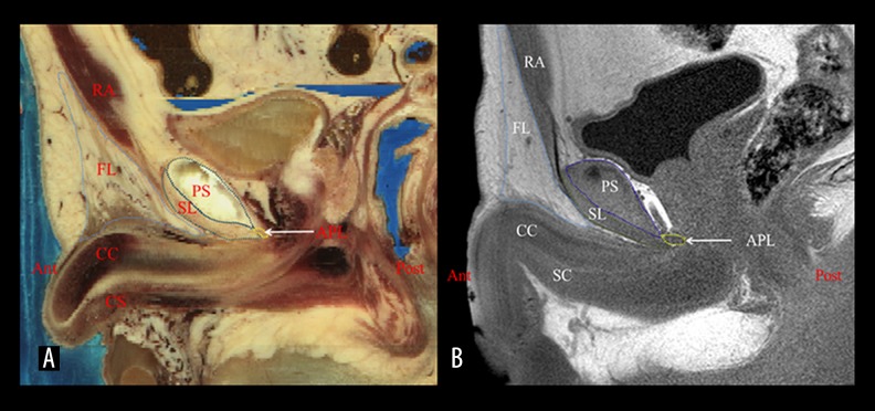
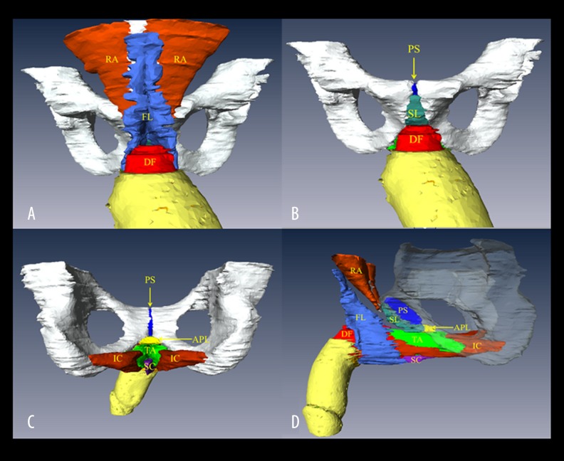
BACKGROUND The aim of this study was to use a three-dimensional (3D) visualization technology to illustrate and describe the anatomical features of the penile suspensory ligamentous system based on the Visible Human data sets and to explore the suspensory mechanism of the penis for the further improvement of the penis-lengthening surgery. MATERIAL AND METHODS Cross-sectional images retrieved from the first Chinese Visible Human (CVH-1), third Chinese Visible Human (CVH-3), and Visible Human Male (VHM) data sets were used to segment the suspensory ligamentous system and its adjacent structures. The magnetic resonance imaging (MRI) images of this system were studied and compared with those from the Visible Human data sets. The 3D models reconstructed from the Visible Human data sets were used to provide morphological features of the penile suspensory ligamentous system and its related structures. RESULTS The fundiform ligament was a superficial, loose, fibro-fatty tissue which originated from Scarpa's fascia superiorly and continued to the scrotal septum inferiorly. The suspensory ligament and arcuate pubic ligament were dense fibrous connective tissues which started from the pubic symphysis and terminated by attaching to the tunica albuginea of the corpora cavernosa. Furthermore, the arcuate pubic ligament attached to the inferior rami of the pubis laterally. CONCLUSIONS The 3D model based on Visible Human data sets can be used to clarify the anatomical features of the suspensory ligamentous system, thereby contributing to the improvement of penis-lengthening surgery.
背景 本研究的目的是利用三维(3D)可视化技术,基于可视人数据集阐明和描述阴茎悬韧带系统的解剖特征,并探索阴茎的悬吊机制,以进一步改进阴茎延长手术。材料与方法 从首个中国可视人(CVH - 1)、第三个中国可视人(CVH - 3)和可视男性(VHM)数据集中检索横断面图像,用于分割悬韧带系统及其相邻结构。研究了该系统的磁共振成像(MRI)图像,并与可视人数据集的图像进行比较。从可视人数据集重建的3D模型用于提供阴茎悬韧带系统及其相关结构的形态特征。结果 耻骨前襞韧带是一种浅表、疏松的纤维脂肪组织,向上起源于Scarpa筋膜,向下延续至阴囊中隔。悬韧带和耻骨弓状韧带是致密的纤维结缔组织,起自耻骨联合,止于阴茎海绵体白膜。此外,耻骨弓状韧带向外附着于耻骨下支。结论 基于可视人数据集的3D模型可用于阐明悬韧带系统的解剖特征,从而有助于改进阴茎延长手术。