• 文献检索
  • 文档翻译
  • 深度研究
  • 学术资讯
  • Suppr Zotero 插件Zotero 插件
  • 邀请有礼
  • 套餐&价格
  • 历史记录
应用&插件
Suppr Zotero 插件Zotero 插件浏览器插件Mac 客户端Windows 客户端微信小程序
定价
高级版会员购买积分包购买API积分包
服务
文献检索文档翻译深度研究API 文档MCP 服务
关于我们
关于 Suppr公司介绍联系我们用户协议隐私条款
关注我们

Suppr 超能文献

核心技术专利:CN118964589B侵权必究
粤ICP备2023148730 号-1Suppr @ 2026

文献检索

告别复杂PubMed语法,用中文像聊天一样搜索,搜遍4000万医学文献。AI智能推荐,让科研检索更轻松。

立即免费搜索

文件翻译

保留排版,准确专业,支持PDF/Word/PPT等文件格式,支持 12+语言互译。

免费翻译文档

深度研究

AI帮你快速写综述,25分钟生成高质量综述,智能提取关键信息,辅助科研写作。

立即免费体验

RUNX3调节缺氧诱导的人心脏微血管内皮细胞的内皮-间充质转化。

RUNX3 modulates hypoxia-induced endothelial-to-mesenchymal transition of human cardiac microvascular endothelial cells.

作者信息

Liu Yanhua, Zou Jin, Li Bingong, Wang Yuqin, Wang Delong, Hao Yanqin, Ke Xuan, Li Xingxing

机构信息

Department of Cardiology, The First Affiliated Hospital, Nanchang University, Nanchang, Jiangxi 330006, P.R. China.

Department of Cardiology, Loudi Central Hospital, Loudi, Hunan 417000, P.R. China.

出版信息

Int J Mol Med. 2017 Jul;40(1):65-74. doi: 10.3892/ijmm.2017.2998. Epub 2017 May 22.

DOI:10.3892/ijmm.2017.2998
PMID:28534977
原文链接:https://pmc.ncbi.nlm.nih.gov/articles/PMC5466396/
Abstract

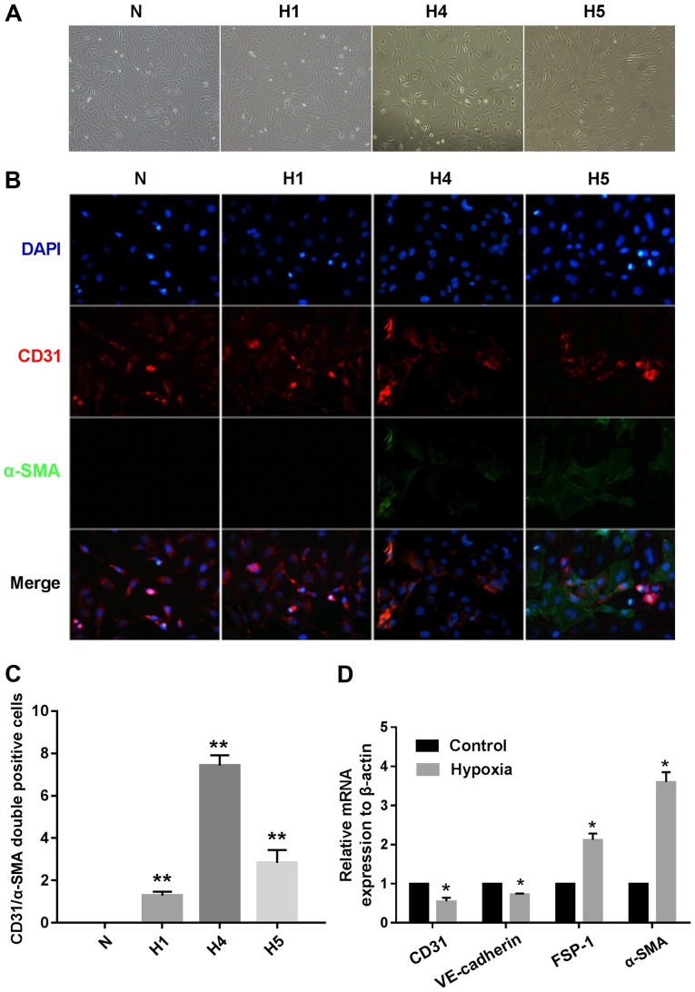
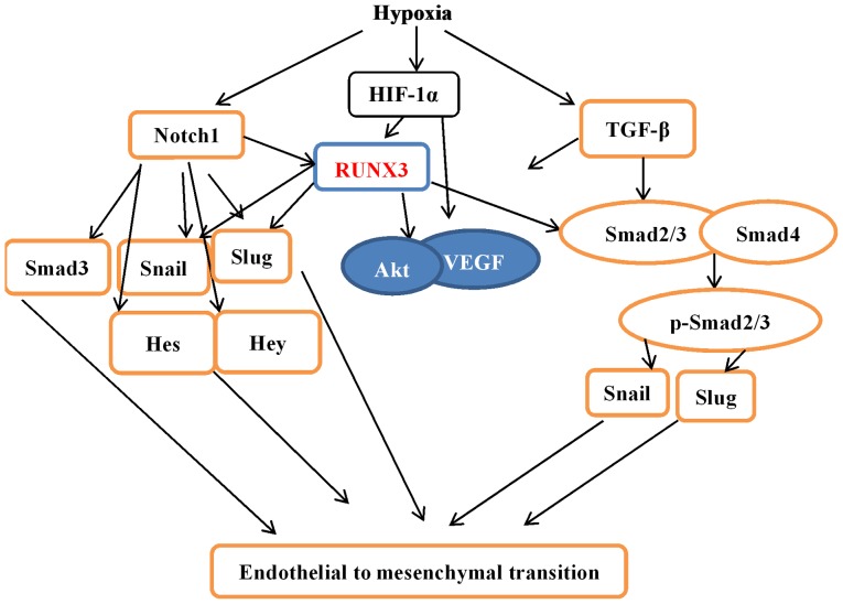
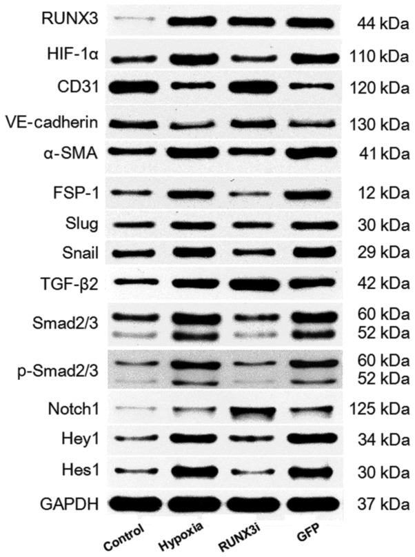
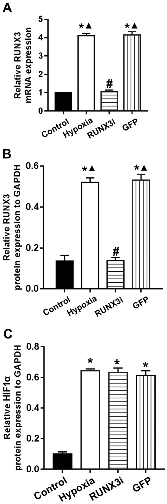
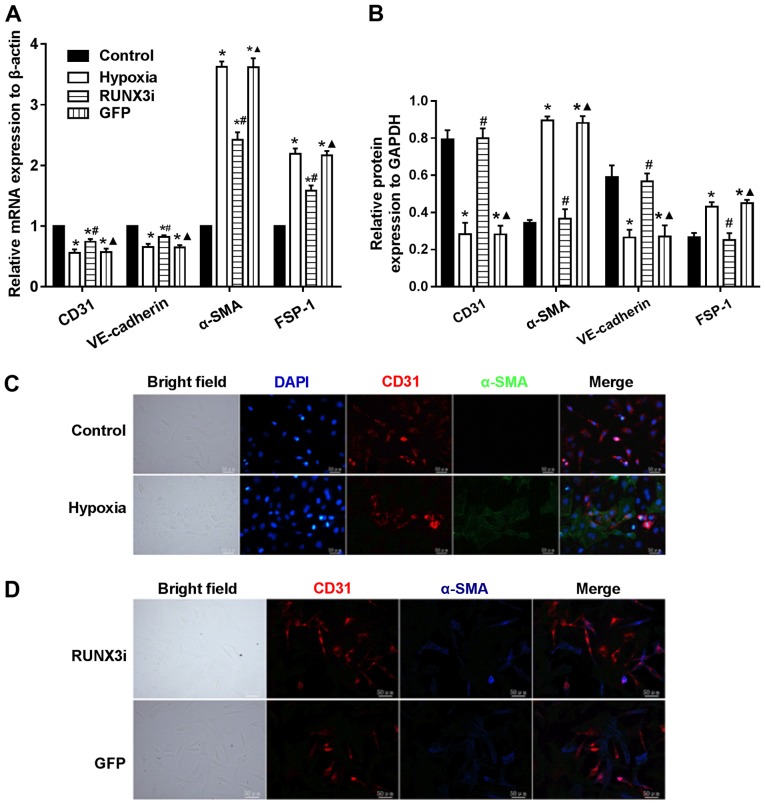
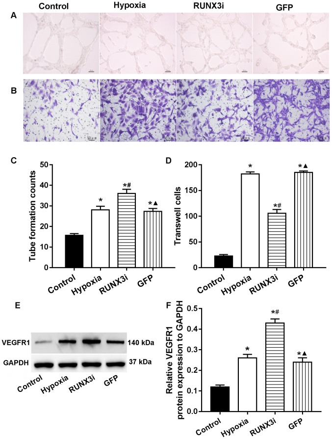
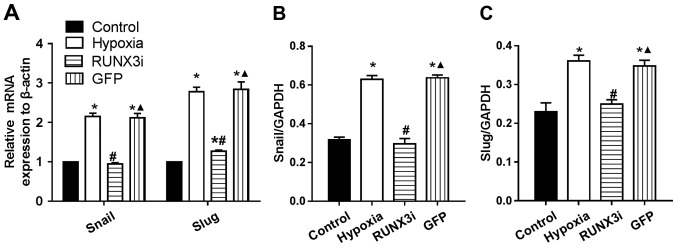
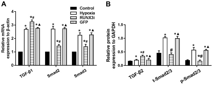
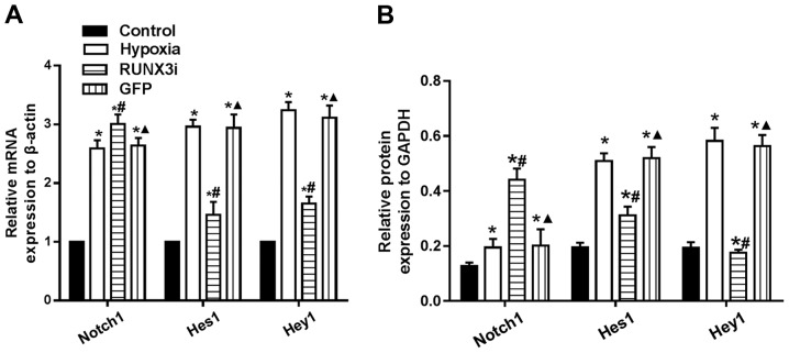
Endothelial-mesenchymal transition (EndMT) is an essential mechanism in the cardiovascular system, for both cardiovascular development and cardiovascular diseases (CVDs). Recent studies indicate that runt-related transcription factor 3 (RUNX3) contributes to EndMT and endothelial cell dysfunction. However, the underlying molecular mechanism remains unknown. The present study was designed to investigate the role of RUNX3 in EndMT and endothelial cell function, and to elucidate the underlying molecular mechanism. Human cardiac microvascular endothelial cells (HCMECs) were incubated in strictly controlled hypoxic conditions (1% O2). HCMECs were cultured under normoxic conditions (21% O2), and then moved to a strictly controlled hypoxic environment (1% O2). Under this hypoxic condition, the cells were transfected with the lentiviral vector containing RUNX3 or an empty lentiviral vector for 8 h. After the cells were cultured under hypoxic conditions for 4 days, CD31 and α-smooth muscle actin colocalization were assessed by immunofluorescence microscopy. Transwell migration and tube formation assays were used to examine the migration and angiogenesis ability. RT-qPCR and western blotting were used to determine the expression of molecules involved in EndMT. Hypoxia induced the transition of HCMECs to mesenchymal cells and markedly promoted tube formation and cell migration. Transforming growth factor-β (TGF-β) and Notch signaling were activated during the hypoxia-induced EndMT of HCMECs. RUNX3 knockdown attenuated EndMT of HCMECs, promoted angiogenic phenotype, and reduced endothelial cell migration. In conclusion, our results showed that RUNX3 knockdown attenuated hypoxia-induced EndMT and reversed endothelial cell functions. RUNX3 is a common downstream target of TGF-β and Notch signaling, and may be a novel therapeutic target for treating CVD mediated by EndMT.

摘要

内皮-间充质转化(EndMT)是心血管系统中一个重要机制,在心血管发育和心血管疾病(CVDs)中均发挥作用。最近的研究表明, runt相关转录因子3(RUNX3)促成EndMT和内皮细胞功能障碍。然而,其潜在分子机制仍不清楚。本研究旨在探讨RUNX3在EndMT和内皮细胞功能中的作用,并阐明其潜在分子机制。将人心脏微血管内皮细胞(HCMECs)置于严格控制的低氧条件(1% O₂)下培养。HCMECs先在常氧条件(21% O₂)下培养,然后转移至严格控制的低氧环境(1% O₂)。在此低氧条件下,用含RUNX3的慢病毒载体或空慢病毒载体转染细胞8小时。细胞在低氧条件下培养4天后,通过免疫荧光显微镜评估CD31和α-平滑肌肌动蛋白的共定位。采用Transwell迁移和管形成实验检测迁移和血管生成能力。RT-qPCR和蛋白质免疫印迹法用于测定EndMT相关分子的表达。低氧诱导HCMECs向间充质细胞转变,并显著促进管形成和细胞迁移。在低氧诱导的HCMECs的EndMT过程中,转化生长因子-β(TGF-β)和Notch信号被激活。RUNX3基因敲低减弱了HCMECs的EndMT,促进了血管生成表型,并减少了内皮细胞迁移。总之,我们的结果表明,RUNX3基因敲低减弱了低氧诱导的EndMT并逆转了内皮细胞功能。RUNX3是TGF-β和Notch信号的共同下游靶点,可能是治疗由EndMT介导的CVD的新治疗靶点。

https://cdn.ncbi.nlm.nih.gov/pmc/blobs/9b1f/5466396/4159eac97961/IJMM-40-01-0065-g08.jpg
https://cdn.ncbi.nlm.nih.gov/pmc/blobs/9b1f/5466396/0ecef6ebe629/IJMM-40-01-0065-g00.jpg
https://cdn.ncbi.nlm.nih.gov/pmc/blobs/9b1f/5466396/788ce5aaa89d/IJMM-40-01-0065-g01.jpg
https://cdn.ncbi.nlm.nih.gov/pmc/blobs/9b1f/5466396/2aad83a1eacb/IJMM-40-01-0065-g02.jpg
https://cdn.ncbi.nlm.nih.gov/pmc/blobs/9b1f/5466396/68f652adb0ce/IJMM-40-01-0065-g03.jpg
https://cdn.ncbi.nlm.nih.gov/pmc/blobs/9b1f/5466396/3c88e28085e4/IJMM-40-01-0065-g04.jpg
https://cdn.ncbi.nlm.nih.gov/pmc/blobs/9b1f/5466396/ac6cc0710d7a/IJMM-40-01-0065-g05.jpg
https://cdn.ncbi.nlm.nih.gov/pmc/blobs/9b1f/5466396/665d2dc59425/IJMM-40-01-0065-g06.jpg
https://cdn.ncbi.nlm.nih.gov/pmc/blobs/9b1f/5466396/23b1d84a95e1/IJMM-40-01-0065-g07.jpg
https://cdn.ncbi.nlm.nih.gov/pmc/blobs/9b1f/5466396/4159eac97961/IJMM-40-01-0065-g08.jpg

相似文献

1
RUNX3 modulates hypoxia-induced endothelial-to-mesenchymal transition of human cardiac microvascular endothelial cells.RUNX3调节缺氧诱导的人心脏微血管内皮细胞的内皮-间充质转化。
Int J Mol Med. 2017 Jul;40(1):65-74. doi: 10.3892/ijmm.2017.2998. Epub 2017 May 22.
2
[Knockdown of RUNX3 inhibits hypoxia-induced endothelial-to-mesenchymal transition of human cardiac microvascular endothelial cells].[RUNX3基因敲低抑制缺氧诱导的人心脏微血管内皮细胞内皮-间充质转化]
Xi Bao Yu Fen Zi Mian Yi Xue Za Zhi. 2016 Dec;32(12):1627-1631.
3
Autophagy attenuates endothelial-to-mesenchymal transition by promoting Snail degradation in human cardiac microvascular endothelial cells.自噬通过促进人心脏微血管内皮细胞中 Snail 的降解来减弱内皮细胞向间充质转化。
Biosci Rep. 2017 Sep 7;37(5). doi: 10.1042/BSR20171049. Print 2017 Oct 31.
4
Protective effects of acetylcholine on hypoxia-induced endothelial-to-mesenchymal transition in human cardiac microvascular endothelial cells.乙酰胆碱对缺氧诱导的人心微血管内皮细胞向间充质转化的保护作用。
Mol Cell Biochem. 2020 Oct;473(1-2):101-110. doi: 10.1007/s11010-020-03811-w. Epub 2020 Jun 29.
5
Downregulation of acylglycerol kinase suppresses high-glucose-induced endothelial-mesenchymal transition in human retinal microvascular endothelial cells through regulating the LPAR1/TGF-β/Notch signaling pathway.酰基甘油激酶下调抑制高糖诱导的人视网膜微血管内皮细胞的内皮-间充质转化通过调节 LPAR1/TGF-β/Notch 信号通路。
Can J Physiol Pharmacol. 2022 Feb;100(2):142-150. doi: 10.1139/cjpp-2021-0265. Epub 2021 Sep 24.
6
The mechanism of TGF-β/miR-155/c-Ski regulates endothelial-mesenchymal transition in human coronary artery endothelial cells.TGF-β/miR-155/c-Ski 调控人冠状动脉内皮细胞内皮-间质转化的机制。
Biosci Rep. 2017 Aug 23;37(4). doi: 10.1042/BSR20160603. Print 2017 Aug 31.
7
RUNX3 maintains the mesenchymal phenotype after termination of the Notch signal.RUNX3 在 Notch 信号终止后维持间充质表型。
J Biol Chem. 2011 Apr 1;286(13):11803-13. doi: 10.1074/jbc.M111.222331. Epub 2011 Feb 2.
8
Tongxinluo Attenuates Myocardiac Fibrosis after Acute Myocardial Infarction in Rats via Inhibition of Endothelial-to-Mesenchymal Transition.通心络通过抑制血管内皮细胞向间充质细胞转化减轻大鼠急性心肌梗死后心肌纤维化。
Biomed Res Int. 2019 Jun 16;2019:6595437. doi: 10.1155/2019/6595437. eCollection 2019.
9
Integrin β3 Mediates the Endothelial-to-Mesenchymal Transition via the Notch Pathway.整合素β3通过Notch信号通路介导内皮-间充质转化
Cell Physiol Biochem. 2018;49(3):985. doi: 10.1159/000493229. Epub 2018 Sep 7.
10
Relaxin inhibits cardiac fibrosis and endothelial-mesenchymal transition via the Notch pathway.松弛素通过Notch信号通路抑制心脏纤维化和内皮-间充质转化。
Drug Des Devel Ther. 2015 Aug 11;9:4599-611. doi: 10.2147/DDDT.S85399. eCollection 2015.

引用本文的文献

1
Study on the mechanism of OSM participating in myocardial fibrosis by inhibiting TGFβ-induced EndMT of cardiac microvascular endothelial cells through SPARC/SMAD signaling.关于通过SPARC/SMAD信号通路抑制转化生长因子β诱导的心脏微血管内皮细胞内皮-间质转化,从而探讨骨形态发生蛋白21参与心肌纤维化机制的研究
Naunyn Schmiedebergs Arch Pharmacol. 2025 Apr;398(4):4479-4489. doi: 10.1007/s00210-024-03472-2. Epub 2024 Nov 4.
2
Elucidating the crosstalk between endothelial-to-mesenchymal transition (EndoMT) and endothelial autophagy in the pathogenesis of atherosclerosis.阐明动脉粥样硬化发病机制中内皮细胞向间充质转化(EndoMT)与内皮细胞自噬之间的串扰。
Vascul Pharmacol. 2024 Jun;155:107368. doi: 10.1016/j.vph.2024.107368. Epub 2024 Mar 26.
3

本文引用的文献

1
Immunoexpression of HIF-1α and VEGF in Periodontal Disease and Healthy Gingival Tissues.低氧诱导因子-1α(HIF-1α)和血管内皮生长因子(VEGF)在牙周病和健康牙龈组织中的免疫表达
Braz Dent J. 2016 Mar-Apr;27(2):117-22. doi: 10.1590/0103-6440201600533.
2
Hypoxia-induced endothelial-mesenchymal transition is associated with RASAL1 promoter hypermethylation in human coronary endothelial cells.缺氧诱导的内皮-间充质转化与人冠状动脉内皮细胞中RASAL1启动子高甲基化有关。
FEBS Lett. 2016 Apr;590(8):1222-33. doi: 10.1002/1873-3468.12158. Epub 2016 Apr 21.
3
A Switch in Akt Isoforms Is Required for Notch-Induced Snail1 Expression and Protection from Cell Death.
A review on regulation of DNA methylation during post-myocardial infarction.
心肌梗死后DNA甲基化调控的综述
Front Pharmacol. 2024 Feb 13;15:1267585. doi: 10.3389/fphar.2024.1267585. eCollection 2024.
4
Tongxinluo Activates PI3K/AKT Signaling Pathway to Inhibit Endothelial Mesenchymal Transition and Attenuate Myocardial Fibrosis after Ischemia-Reperfusion in Mice.通心络通过激活 PI3K/AKT 信号通路抑制缺血再灌注后小鼠内皮间质转化并减轻心肌纤维化。
Chin J Integr Med. 2024 Jul;30(7):608-615. doi: 10.1007/s11655-024-3652-5. Epub 2024 Feb 22.
5
Liver X receptor alpha ensures blood-brain barrier function by suppressing SNAI2.肝 X 受体α通过抑制 SNAI2 来确保血脑屏障功能。
Cell Death Dis. 2023 Nov 28;14(11):781. doi: 10.1038/s41419-023-06316-8.
6
miR-221-3p targets Ang-2 to inhibit the transformation of HCMECs to tip cells.miR-221-3p 通过靶向 Ang-2 抑制 HCMECs 向 Tip 细胞的转化。
J Cell Mol Med. 2023 Nov;27(21):3247-3258. doi: 10.1111/jcmm.17892. Epub 2023 Jul 31.
7
Transcriptomic Context of Expression in Monocytes: A Cross-Sectional Analysis.单核细胞中表达的转录组学背景:一项横断面分析。
Biomedicines. 2023 Jun 13;11(6):1698. doi: 10.3390/biomedicines11061698.
8
LncRNA LINC00961 regulates endothelial‑mesenchymal transition via the PTEN‑PI3K‑AKT pathway.长链非编码 RNA LINC00961 通过 PTEN-PI3K-AKT 通路调节血管内皮-间质转化。
Mol Med Rep. 2022 Jul;26(1). doi: 10.3892/mmr.2022.12762. Epub 2022 Jun 3.
9
Yes-associated protein promotes endothelial-to-mesenchymal transition of endothelial cells in choroidal neovascularization fibrosis.Yes相关蛋白促进脉络膜新生血管纤维化中内皮细胞的内皮-间充质转化。
Int J Ophthalmol. 2022 May 18;15(5):701-710. doi: 10.18240/ijo.2022.05.03. eCollection 2022.
10
Establishing a competing endogenous RNA (ceRNA)-immunoregulatory network associated with the progression of Alzheimer's disease.建立与阿尔茨海默病进展相关的竞争性内源性RNA(ceRNA)免疫调节网络。
Ann Transl Med. 2022 Jan;10(2):65. doi: 10.21037/atm-21-6762..
Notch诱导的Snail1表达及细胞死亡保护需要Akt亚型的转换。
Mol Cell Biol. 2015 Dec 28;36(6):923-40. doi: 10.1128/MCB.01074-15.
4
The emerging role of RUNX3 in cancer metastasis (Review).RUNX3在癌症转移中的新作用(综述)。
Oncol Rep. 2016 Mar;35(3):1227-36. doi: 10.3892/or.2015.4515. Epub 2015 Dec 24.
5
Relaxin inhibits cardiac fibrosis and endothelial-mesenchymal transition via the Notch pathway.松弛素通过Notch信号通路抑制心脏纤维化和内皮-间充质转化。
Drug Des Devel Ther. 2015 Aug 11;9:4599-611. doi: 10.2147/DDDT.S85399. eCollection 2015.
6
Protective effects of Notch1 signaling activation against high glucose-induced myocardial cell injury: Analysis of its mechanisms of action.Notch1信号激活对高糖诱导的心肌细胞损伤的保护作用:作用机制分析
Int J Mol Med. 2015 Sep;36(3):897-903. doi: 10.3892/ijmm.2015.2294. Epub 2015 Jul 22.
7
Snail Is a Direct Target of Hypoxia-inducible Factor 1α (HIF1α) in Hypoxia-induced Endothelial to Mesenchymal Transition of Human Coronary Endothelial Cells.在缺氧诱导的人冠状动脉内皮细胞向间充质细胞转变过程中,Snail是缺氧诱导因子1α(HIF1α)的直接靶点。
J Biol Chem. 2015 Jul 3;290(27):16653-64. doi: 10.1074/jbc.M115.636944. Epub 2015 May 13.
8
Endothelial-to-mesenchymal transition in pulmonary hypertension.肺高血压中的内皮细胞-间充质转化。
Circulation. 2015 Mar 17;131(11):1006-18. doi: 10.1161/CIRCULATIONAHA.114.008750. Epub 2015 Jan 15.
9
Signaling mechanisms of the epithelial-mesenchymal transition.上皮-间质转化的信号传导机制
Sci Signal. 2014 Sep 23;7(344):re8. doi: 10.1126/scisignal.2005189.
10
Constitutively active Notch1 signaling promotes endothelial‑mesenchymal transition in a conditional transgenic mouse model.在条件性转基因小鼠模型中,组成型激活的Notch1信号传导促进内皮-间充质转化。
Int J Mol Med. 2014 Sep;34(3):669-76. doi: 10.3892/ijmm.2014.1818. Epub 2014 Jun 24.