Agwuna K K, Eze C U, Ukoha P O, Umeh U A
Department of Radiation Medicine, University of Nigeria, Enugu Campus, Enugu, Nigeria.
Department of Medical Radiography and Radiological Sciences, Faculty of Health Sciences and Technology, College of Medicine, University of , Enugu Campus, Enugu, Nigeria.
Ann Med Health Sci Res. 2016 Nov-Dec;6(6):335-340. doi: 10.4103/amhsr.amhsr_457_15.
The accuracy of common ultrasound parameters for the estimation of gestational age (GA) decreases as pregnancy advances in age. Hence, there is need to explore other parameters that may complement the established fetal biometric parameters in predicting GA in late pregnancy.
The aim of this study is to determine the relationship between the sonographic placental thickness (PT) and GA in the second and third trimesters.
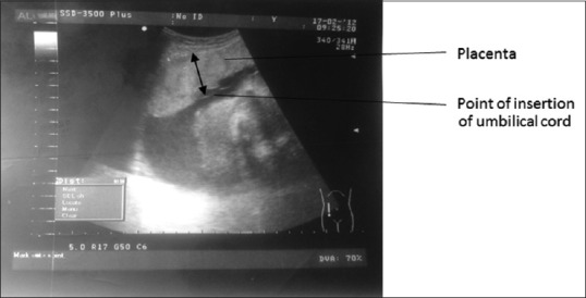
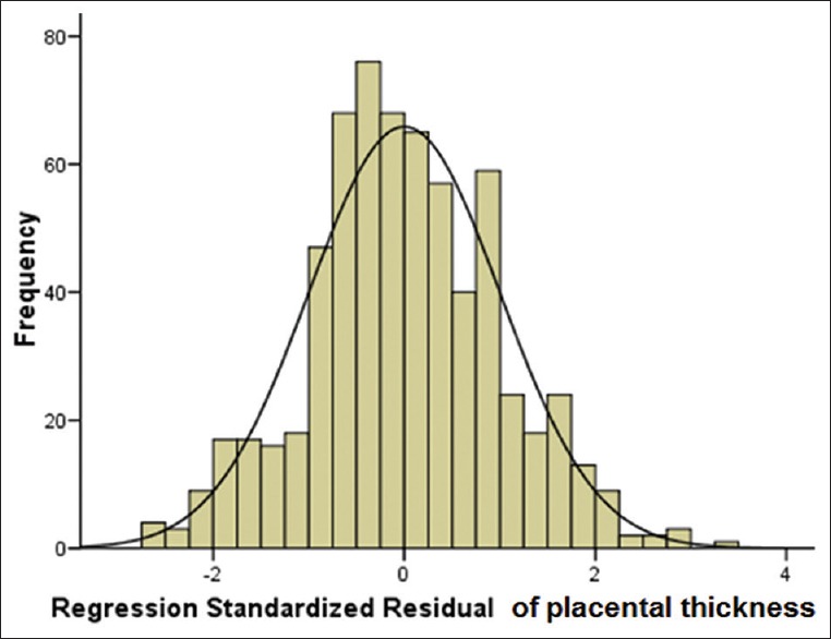
A cross-sectional study of 627 normal pregnant women with GA between 14 and 40 weeks was conducted at the University of Nigeria Teaching Hospital Ituku-Ozalla, Enugu from May 2013 to February 2014 by sonography. Anteroposterior diameter of the placenta was measured at the level of the umbilical cord insertion. The last menstrual period of the women, femur length, biparietal diameter, head circumference, and abdominal circumference of the fetus were measured for GA estimation. Descriptive statistics, regression analysis, and independent sample -test were used in statistical analysis.
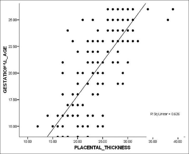
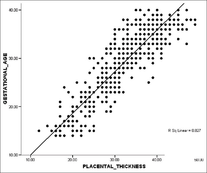
Mean PT was 23.2 (2.8) mm in the second trimester and 36.1 (3.6) mm in the third trimester. There was a significant difference between the values in the present study and values from similar studies in other populations ( < 0.04). There was a strong relationship between GA and PT and the following mathematical relationships for the second and third trimesters were obtained in the GA = 0.982 (PT) + 3.614 and GA = 0.977 (PT) + 3.354, respectively.
Population-specific charts for PT may be used to estimate GA in the second and third trimesters.
随着孕周增加,用于估算孕周(GA)的常见超声参数的准确性会降低。因此,有必要探索其他参数,以在预测晚期妊娠的孕周时补充已有的胎儿生物测量参数。
本研究的目的是确定孕中期和孕晚期超声测量的胎盘厚度(PT)与孕周之间的关系。
2013年5月至2014年2月,在尼日利亚大学教学医院伊图库-奥扎拉(位于埃努古),对627名孕周在14至40周之间的正常孕妇进行了一项横断面超声研究。在脐带插入处测量胎盘的前后径。测量孕妇的末次月经时间、胎儿的股骨长度、双顶径、头围和腹围,以估算孕周。统计分析采用描述性统计、回归分析和独立样本t检验。
孕中期平均胎盘厚度为23.2(2.8)mm,孕晚期为36.1(3.6)mm。本研究中的值与其他人群类似研究中的值之间存在显著差异(P<0.04)。孕周与胎盘厚度之间存在密切关系,孕中期和孕晚期分别得到以下数学关系:GA = 0.982(PT)+ 3.614和GA = 0.977(PT)+ 3.354。
特定人群的胎盘厚度图表可用于估算孕中期和孕晚期的孕周。