Centre for Topical Drug Delivery and Toxicology School of Life and Medical Sciences, University of Hertfordshire, Hatfield, Hertfordshire, AL10 9AB, UK.
Department of Pharmaceutical Biochemistry and Molecular Diagnostics Pharmacy Faculty, Medical University of Lodz, 1 Muszynskiego Street, 90-151, Lodz, Poland.
Pharm Res. 2017 Dec;34(12):2466-2476. doi: 10.1007/s11095-017-2176-5. Epub 2017 May 24.
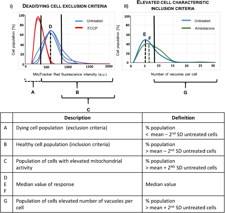
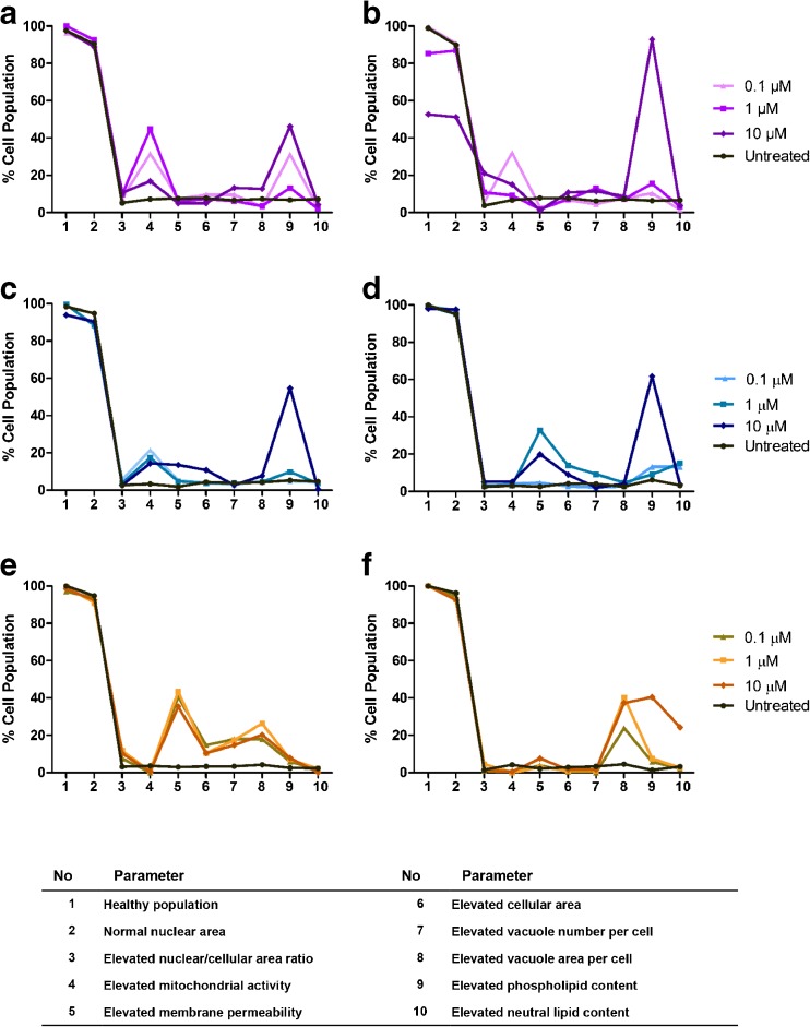
Progress to the clinic may be delayed or prevented when vacuolated or "foamy" alveolar macrophages are observed during non-clinical inhalation toxicology assessment. The first step in developing methods to study this response in vitro is to characterize macrophage cell lines and their response to drug exposures.
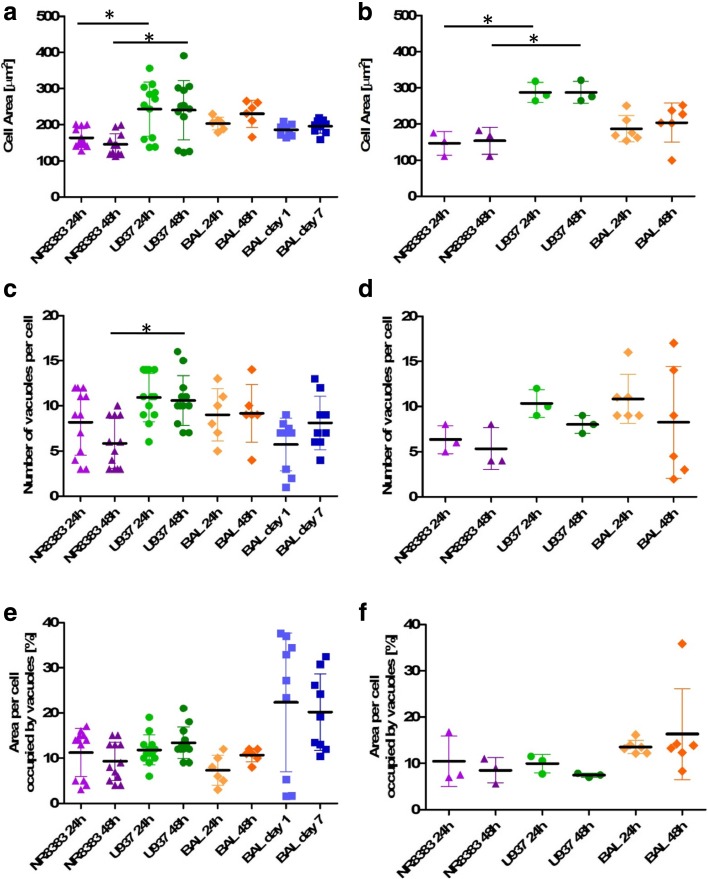
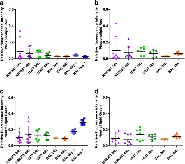
Human (U937) and rat (NR8383) cell lines and primary rat alveolar macrophages obtained by bronchoalveolar lavage were characterized using high content fluorescence imaging analysis quantification of cell viability, morphometry, and phospholipid and neutral lipid accumulation.
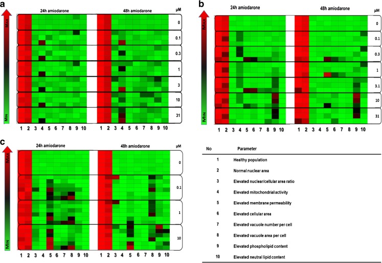
Cell health, morphology and lipid content were comparable (p < 0.05) for both cell lines and the primary macrophages in terms of vacuole number, size and lipid content. Responses to amiodarone, a known inducer of phospholipidosis, required analysis of shifts in cell population profiles (the proportion of cells with elevated vacuolation or lipid content) rather than average population data which was insensitive to the changes observed.
A high content image analysis assay was developed and used to provide detailed morphological characterization of rat and human alveolar-like macrophages and their response to a phospholipidosis-inducing agent. This provides a basis for development of assays to predict or understand macrophage vacuolation following inhaled drug exposure.
在非临床吸入毒理学评估中观察到空泡状或“泡沫状”肺泡巨噬细胞时,可能会延迟或阻止其进入临床阶段。开发体外研究这种反应的方法的第一步是对巨噬细胞系及其对药物暴露的反应进行特征描述。
使用高内涵荧光成像分析对人(U937)和大鼠(NR8383)细胞系以及通过支气管肺泡灌洗获得的原代大鼠肺泡巨噬细胞进行特征描述,定量分析细胞活力、形态计量学以及磷脂和中性脂质的积累。
就空泡数量、大小和脂质含量而言,两种细胞系和原代巨噬细胞的细胞健康状况、形态和脂质含量相当(p<0.05)。对于已知会诱导磷脂蓄积的胺碘酮,需要分析细胞群体分布的变化(具有较高空泡化或脂质含量的细胞比例),而不是对观察到的变化不敏感的平均群体数据。
开发了一种高内涵图像分析测定法,用于对大鼠和人肺泡样巨噬细胞及其对磷脂蓄积诱导剂的反应进行详细的形态特征描述。这为开发预测或了解吸入药物暴露后巨噬细胞空泡化的测定法提供了基础。