Department of Immunology, School of Basic Medical Sciences, Key Laboratory of Medical Immunology of Ministry of Public Health, Peking University Health Science Center, Beijing, China.
Institute of Biological Sciences, Jinzhou Medical University, Jinzhou, Liaoning, China.
Sci Rep. 2017 May 25;7(1):2421. doi: 10.1038/s41598-017-02653-9.
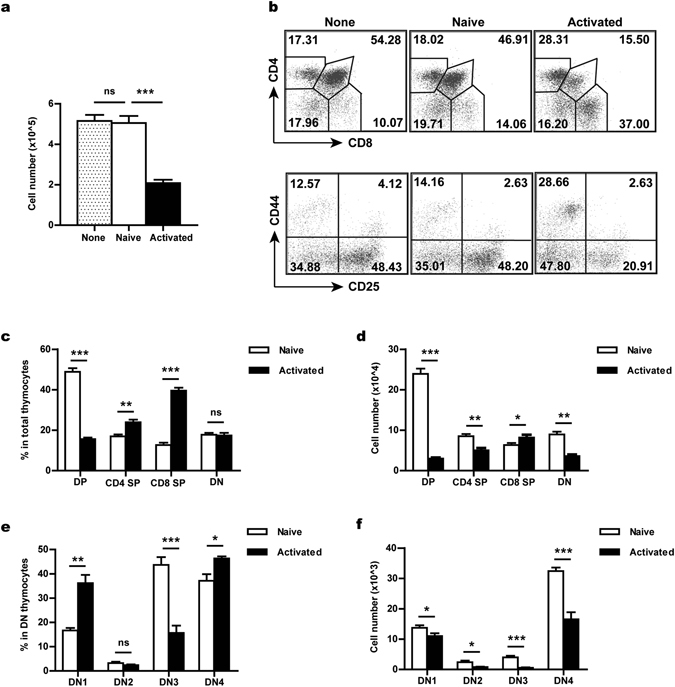
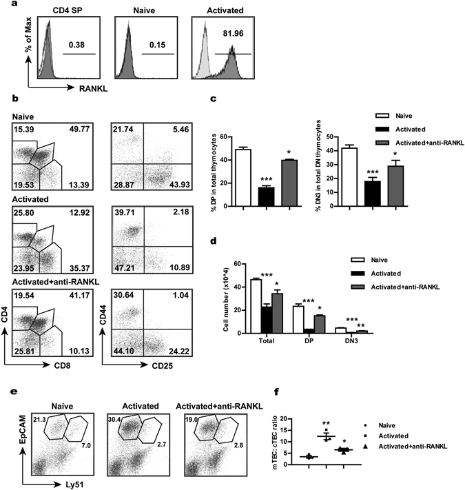
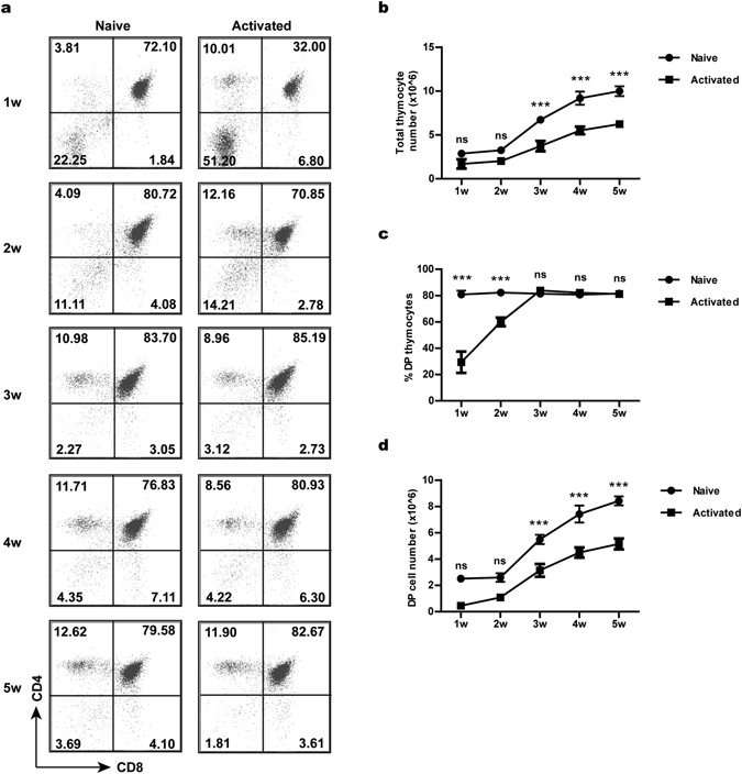
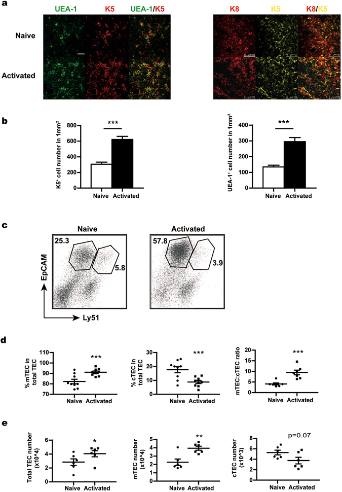
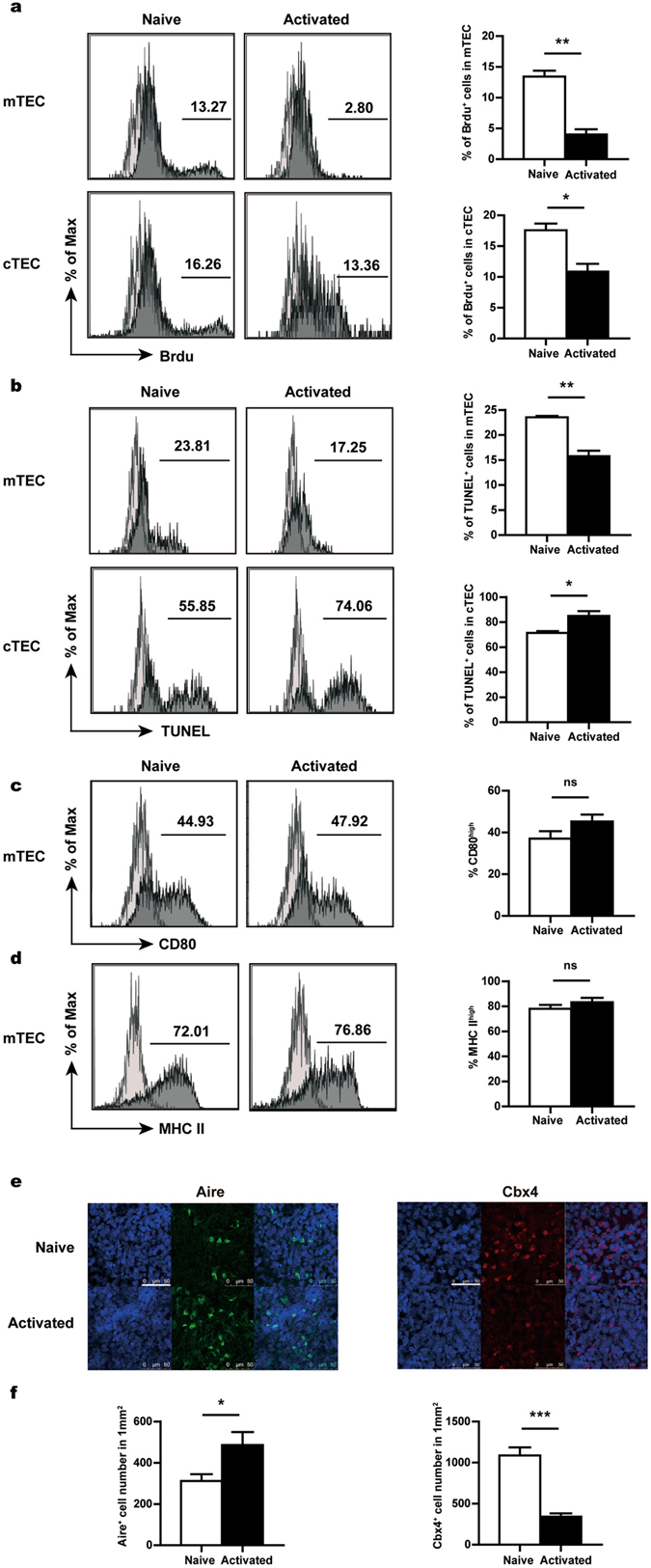
Activated T cells have been shown to be able to recirculate into the thymus from the periphery. The present study was aimed to elucidate the functional consequences of thymic homing of activated T cells upon developing thymocytes and thymic epithelial cells (TEC). In the presence of activated T cells, especially CD4 T cells, T cell development was found to be inhibited in thymic organ cultures with markedly reduced cellularity. Thymic transplantation demonstrated that the inhibitory effect was most likely due to a defective microenvironment. As the major component of the thymic stroma, the TEC compartment was severely disturbed after prolonged exposure to the activated T cells. In addition to reduced cell proliferation, TEC differentiation was heavily skewed to the mTEC lineage. Furthermore, we demonstrated that RANKL highly expressed by activated CD4 T cells was primarily responsible for the detrimental effects. Presumably, excessive RANK signaling drove overproduction of mTECs and possibly exhaustion of epithelial progenitors, thereby facilitating the deterioration of the epithelial structures. These findings not only reveal a novel activity of activated T cells re-entering the thymus, but also provide a new perspective for understanding the mechanism underlying thymic involution.
已证实激活的 T 细胞能够从外周组织再循环进入胸腺。本研究旨在阐明激活的 T 细胞归巢至胸腺对胸腺细胞和胸腺上皮细胞(TEC)的功能影响。在存在激活的 T 细胞,特别是 CD4 T 细胞的情况下,发现胸腺器官培养中的 T 细胞发育受到抑制,细胞数量明显减少。胸腺移植表明,这种抑制作用很可能是由于微环境缺陷所致。作为胸腺基质的主要成分,TEC 区室在长时间暴露于激活的 T 细胞后受到严重干扰。除了细胞增殖减少外,TEC 分化严重偏向 mTEC 谱系。此外,我们证明激活的 CD4 T 细胞高表达的 RANKL 是造成这种损害的主要原因。推测过度的 RANK 信号传导导致 mTEC 的过度产生,并且可能耗尽上皮祖细胞,从而促进上皮结构的恶化。这些发现不仅揭示了激活的 T 细胞重新进入胸腺的新活性,而且为理解胸腺萎缩的机制提供了新的视角。