• 文献检索
  • 文档翻译
  • 深度研究
  • 学术资讯
  • Suppr Zotero 插件Zotero 插件
  • 邀请有礼
  • 套餐&价格
  • 历史记录
应用&插件
Suppr Zotero 插件Zotero 插件浏览器插件Mac 客户端Windows 客户端微信小程序
定价
高级版会员购买积分包购买API积分包
服务
文献检索文档翻译深度研究API 文档MCP 服务
关于我们
关于 Suppr公司介绍联系我们用户协议隐私条款
关注我们

Suppr 超能文献

核心技术专利:CN118964589B侵权必究
粤ICP备2023148730 号-1Suppr @ 2026

文献检索

告别复杂PubMed语法,用中文像聊天一样搜索,搜遍4000万医学文献。AI智能推荐,让科研检索更轻松。

立即免费搜索

文件翻译

保留排版,准确专业,支持PDF/Word/PPT等文件格式,支持 12+语言互译。

免费翻译文档

深度研究

AI帮你快速写综述,25分钟生成高质量综述,智能提取关键信息,辅助科研写作。

立即免费体验

使用定量透射(QT)超声对乳腺囊肿与实性病变诊断的准确性

Accuracy of Cyst Versus Solid Diagnosis in the Breast Using Quantitative Transmission (QT) Ultrasound.

作者信息

Iuanow Elaine, Smith Kathleen, Obuchowski Nancy A, Bullen Jennifer, Klock John C

机构信息

QT Ultrasound Labs, 3 Hamilton Landing Suite 160, Novato, CA 94949.

Department of Quantitative Health Sciences, Cleveland Clinic Foundation, Cleveland, Ohio.

出版信息

Acad Radiol. 2017 Sep;24(9):1148-1153. doi: 10.1016/j.acra.2017.03.024. Epub 2017 May 23.

DOI:10.1016/j.acra.2017.03.024
PMID:28549870
原文链接:https://pmc.ncbi.nlm.nih.gov/articles/PMC5557662/
Abstract

RATIONAL AND OBJECTIVES

This study aims to evaluate the diagnostic utility of breast imaging using transmission ultrasound. We present readers' accuracy in determining whether a breast lesion is a cyst versus a solid using transmission ultrasound as an adjunct to mammography.

MATERIALS AND METHODS

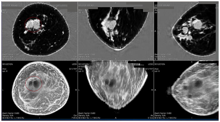
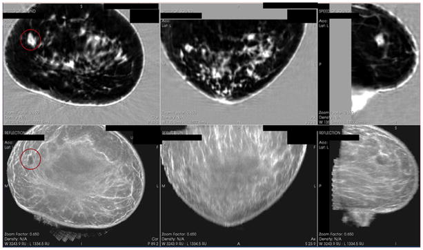
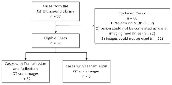
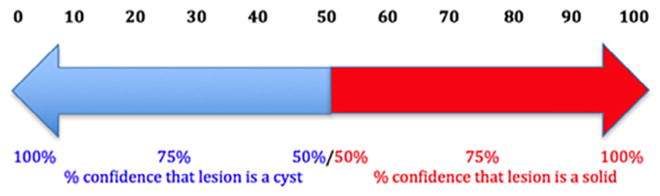
This retrospective multi-reader, multi-case receiver operating characteristic study included 37 lesions seen on mammography and transmission ultrasound. Cyst cases were confirmed as cysts using their appearance on handheld ultrasound. Solid cases were confirmed as solids with pathology results. Fourteen readers performed blinded, randomized reads with mammography + quantitative transmission scan images, assigning both a confidence score (0-100) and a binary classification of cyst or solid. A 95% percentile bootstrap confidence interval (CI) was computed for the readers' mean receiver operating characteristic area, sensitivity, and specificity.

RESULTS

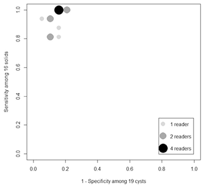
Using the readers' binary classification of cyst or solid lesions, the mean sensitivity and specificity were 0.933 [95% CI: 0.837, 0.995] and 0.858 [95% CI: 0.701, 0.985], respectively. When the readers' confidence scores were used to distinguish a cyst versus solid, the mean receiver operating characteristic area was 0.920 [95% CI: 0.827, 0.985].

CONCLUSIONS

Transmission ultrasound can provide an accurate assessment of a cyst versus a solid lesion in the breast. Prospective clinical trials will further delineate the role of transmission ultrasound as an adjunct to mammography to increase specificity in breast evaluation.

摘要

研究目的与目标

本研究旨在评估透射超声在乳腺成像中的诊断效用。我们展示了读者在使用透射超声作为乳腺X线摄影辅助手段来判断乳腺病变是囊肿还是实性病变时的准确性。

材料与方法

这项回顾性多读者、多病例的接收器操作特性研究纳入了37例在乳腺X线摄影和透射超声检查中发现的病变。囊肿病例通过手持超声检查的表现确诊为囊肿。实性病例通过病理结果确诊为实性病变。14名读者对乳腺X线摄影 + 定量透射扫描图像进行了盲法、随机阅读,给出一个置信度评分(0 - 100)以及囊肿或实性的二元分类。计算了读者平均接收器操作特性曲线下面积、敏感性和特异性的95%百分位数自助置信区间(CI)。

结果

根据读者对囊肿或实性病变的二元分类,平均敏感性和特异性分别为0.933 [95% CI:0.837, 0.995] 和0.858 [95% CI:0.701, 0.985]。当使用读者的置信度评分来区分囊肿和实性病变时,平均接收器操作特性曲线下面积为0.920 [95% CI:0.827, 0.985]。

结论

透射超声能够对乳腺中的囊肿和实性病变进行准确评估。前瞻性临床试验将进一步阐明透射超声作为乳腺X线摄影辅助手段在提高乳腺评估特异性方面的作用。

https://cdn.ncbi.nlm.nih.gov/pmc/blobs/b1ac/5557662/f5d9330fde2a/nihms879252f5.jpg
https://cdn.ncbi.nlm.nih.gov/pmc/blobs/b1ac/5557662/f0952ec949e9/nihms879252f1.jpg
https://cdn.ncbi.nlm.nih.gov/pmc/blobs/b1ac/5557662/a1fe86b0aa2d/nihms879252f2.jpg
https://cdn.ncbi.nlm.nih.gov/pmc/blobs/b1ac/5557662/719365b91e4b/nihms879252f3.jpg
https://cdn.ncbi.nlm.nih.gov/pmc/blobs/b1ac/5557662/bbe6cae83fe0/nihms879252f4.jpg
https://cdn.ncbi.nlm.nih.gov/pmc/blobs/b1ac/5557662/f5d9330fde2a/nihms879252f5.jpg

相似文献

1
Accuracy of Cyst Versus Solid Diagnosis in the Breast Using Quantitative Transmission (QT) Ultrasound.使用定量透射(QT)超声对乳腺囊肿与实性病变诊断的准确性
Acad Radiol. 2017 Sep;24(9):1148-1153. doi: 10.1016/j.acra.2017.03.024. Epub 2017 May 23.
2
An Exploratory Multi-reader, Multi-case Study Comparing Transmission Ultrasound to Mammography on Recall Rates and Detection Rates for Breast Cancer Lesions.探索性多读者、多病例研究比较了超声传输与乳腺 X 线摄影对乳腺癌病变的召回率和检出率。
Acad Radiol. 2022 Jan;29 Suppl 1:S10-S18. doi: 10.1016/j.acra.2020.11.011. Epub 2020 Dec 3.
3
3D quantitative breast ultrasound analysis for differentiating fibroadenomas and carcinomas smaller than 1cm.用于鉴别小于1cm的纤维腺瘤和癌的三维定量乳腺超声分析
Eur J Radiol. 2017 Mar;88:141-147. doi: 10.1016/j.ejrad.2017.01.006. Epub 2017 Jan 8.
4
Breast lesion detection and classification: comparison of screen-film mammography and full-field digital mammography with soft-copy reading--observer performance study.乳腺病变的检测与分类:屏-片乳腺摄影与软读片全视野数字化乳腺摄影的比较——观察者性能研究
Radiology. 2005 Oct;237(1):37-44. doi: 10.1148/radiol.2371041605. Epub 2005 Aug 11.
5
Characterization of Cystic Lesions by Spectral Mammography: Results of a Clinical Pilot Study.超声光散射断层成像技术对囊性病灶的特征分析:一项临床初步研究结果
Invest Radiol. 2016 May;51(5):340-7. doi: 10.1097/RLI.0000000000000246.
6
Clinical performance of Siemens digital breast tomosynthesis versus standard supplementary mammography for the assessment of screen-detected soft-tissue abnormalities: a multi-reader study.西门子数字乳腺断层合成技术与标准补充乳腺摄影术在评估筛查发现的软组织异常方面的临床性能:一项多阅片者研究。
Clin Radiol. 2017 Jan;72(1):95.e9-95.e15. doi: 10.1016/j.crad.2016.08.011. Epub 2016 Oct 10.
7
Comparison of radiologist performance with photon-counting full-field digital mammography to conventional full-field digital mammography.与传统全数字乳腺摄影相比,光子计数全数字乳腺摄影的放射科医生性能比较。
Acad Radiol. 2012 Aug;19(8):916-22. doi: 10.1016/j.acra.2012.03.005. Epub 2012 Apr 24.
8
Automated Breast Ultrasound in Breast Cancer Screening of Women With Dense Breasts: Reader Study of Mammography-Negative and Mammography-Positive Cancers.乳腺致密女性乳腺癌筛查中的自动乳腺超声:乳腺X线摄影阴性和阳性癌症的阅片者研究
AJR Am J Roentgenol. 2016 Jun;206(6):1341-50. doi: 10.2214/AJR.15.15367. Epub 2016 Apr 4.
9
Use of single-view digital breast tomosynthesis (DBT) and ultrasound vs. additional views and ultrasound for the assessment of screen-detected abnormalities: German multi-reader study.使用单视图数字乳腺断层合成(DBT)和超声与额外视图及超声用于评估筛查发现的异常情况:德国多阅片者研究
Acta Radiol. 2018 Jul;59(7):782-788. doi: 10.1177/0284185117732600. Epub 2017 Sep 20.
10
The influence of knowledge of mammography findings on the accuracy of breast ultrasound in symptomatic women.乳腺钼靶检查结果的知晓情况对有症状女性乳腺超声检查准确性的影响。
Breast J. 2005 May-Jun;11(3):167-72. doi: 10.1111/j.1075-122X.2005.21643.x.

引用本文的文献

1
Sizing It Up: Concordance between Breast Imaging and Pathologically Determined Tumor Measurement.评估其大小:乳腺影像与病理确定的肿瘤测量之间的一致性。
Ann Surg Oncol. 2025 Jun 16. doi: 10.1245/s10434-025-17663-5.
2
Acoustomotive diffuse correlation spectroscopy for sensing mechanical stiffness in tissue-mimicking phantoms.用于检测仿组织体模中机械硬度的声动力扩散相关光谱技术。
Biomed Opt Express. 2024 Aug 19;15(9):5328-5348. doi: 10.1364/BOE.531963. eCollection 2024 Sep 1.
3
Breast Glandular and Ductal Volume Changes during the Menstrual Cycle: A Study in 48 Breasts Using Ultralow-Frequency Transmitted Ultrasound Tomography/Volography.

本文引用的文献

1
Objective breast tissue image classification using Quantitative Transmission ultrasound tomography.基于定量超声层析成像的乳腺组织图像分类
Sci Rep. 2016 Dec 9;6:38857. doi: 10.1038/srep38857.
2
Anatomy-Correlated Breast Imaging and Visual Grading Analysis Using Quantitative Transmission Ultrasound.使用定量透射超声的解剖学相关乳腺成像与视觉分级分析
Int J Biomed Imaging. 2016;2016:7570406. doi: 10.1155/2016/7570406. Epub 2016 Sep 26.
3
Imaging Performance of Quantitative Transmission Ultrasound.定量透射超声的成像性能
经超低频发射超声断层摄影/体层摄影术研究 48 例乳房,了解月经周期中乳腺腺泡和导管体积的变化。
Tomography. 2024 May 19;10(5):789-805. doi: 10.3390/tomography10050060.
4
Breast Tomographic Ultrasound: The Spectrum from Current Dense Breast Cancer Screenings to Future Theranostic Treatments.乳腺断层超声:从当前致密型乳腺癌筛查到未来的治疗应用。
Tomography. 2024 Apr 15;10(4):554-573. doi: 10.3390/tomography10040044.
5
Low frequency 3D transmission ultrasound tomography: technical details and clinical implications.低频三维传输超声断层成像:技术细节与临床应用
Z Med Phys. 2023 Aug;33(3):427-443. doi: 10.1016/j.zemedi.2023.04.006. Epub 2023 Jun 7.
6
Coherence Metrics for Reader-Independent Differentiation of Cystic From Solid Breast Masses in Ultrasound Images.用于在超声图像中对囊性和实性乳腺肿块进行读者独立区分的相干性度量。
Ultrasound Med Biol. 2023 Jan;49(1):256-268. doi: 10.1016/j.ultrasmedbio.2022.08.018. Epub 2022 Nov 1.
7
The Potential Role of the Fat-Glandular Interface (FGI) in Breast Carcinogenesis: Results from an Ultrasound Tomography (UST) Study.脂肪-腺体界面(FGI)在乳腺癌发生中的潜在作用:超声断层扫描(UST)研究结果
J Clin Med. 2021 Nov 29;10(23):5615. doi: 10.3390/jcm10235615.
8
Multicenter Study of Whole Breast Stiffness Imaging by Ultrasound Tomography (SoftVue) for Characterization of Breast Tissues and Masses.超声断层扫描(SoftVue)对乳腺组织和肿块进行特征化的全乳硬度成像多中心研究。
J Clin Med. 2021 Nov 25;10(23):5528. doi: 10.3390/jcm10235528.
9
Speed-of-sound imaging using diverging waves.利用发散波进行声速成象。
Int J Comput Assist Radiol Surg. 2021 Jul;16(7):1201-1211. doi: 10.1007/s11548-021-02426-w. Epub 2021 Jun 23.
10
Breast Cyst Fluid Analysis Correlations with Speed of Sound Using Transmission Ultrasound.乳腺囊肿液分析与超声透射声速的相关性。
Acad Radiol. 2019 Jan;26(1):76-85. doi: 10.1016/j.acra.2018.03.027. Epub 2018 Jun 7.
Int J Biomed Imaging. 2015;2015:454028. doi: 10.1155/2015/454028. Epub 2015 Oct 28.
4
National expenditure for false-positive mammograms and breast cancer overdiagnoses estimated at $4 billion a year.每年因乳腺钼靶检查假阳性和乳腺癌过度诊断产生的国家支出估计为40亿美元。
Health Aff (Millwood). 2015 Apr;34(4):576-83. doi: 10.1377/hlthaff.2014.1087.
5
Consequences of false-positive screening mammograms.假阳性筛查性乳房 X 光检查的后果。
JAMA Intern Med. 2014 Jun;174(6):954-61. doi: 10.1001/jamainternmed.2014.981.
6
Long-term psychosocial consequences of false-positive screening mammography.假阳性筛查性乳房 X 光检查的长期心理社会后果。
Ann Fam Med. 2013 Mar-Apr;11(2):106-15. doi: 10.1370/afm.1466.
7
Quantitative volumetric breast imaging with 3D inverse scatter computed tomography.采用三维逆散射计算机断层扫描的定量乳腺容积成像。
Annu Int Conf IEEE Eng Med Biol Soc. 2012;2012:1110-3. doi: 10.1109/EMBC.2012.6346129.
8
Non-linear inverse scattering: high resolution quantitative breast tissue tomography.非线性逆散射:高分辨率定量乳腺组织层析成像。
J Acoust Soc Am. 2012 May;131(5):3802-13. doi: 10.1121/1.3699240.
9
Cystic breast lesions: sonographic findings and clinical management.囊性乳腺病变:超声表现与临床处理。
J Ultrasound Med. 2010 Nov;29(11):1617-26. doi: 10.7863/jum.2010.29.11.1617.
10
Cystic breast masses and the ACRIN 6666 experience.乳腺囊性肿块与ACRIN 6666研究经验
Radiol Clin North Am. 2010 Sep;48(5):931-87. doi: 10.1016/j.rcl.2010.06.007.