Translational and Molecular Imaging Institute, Icahn School of Medicine at Mount Sinai, New York, NY, United States.
Department of Radiology, Icahn School of Medicine at Mount Sinai, New York, NY, United States.
Sci Rep. 2017 May 26;7(1):2452. doi: 10.1038/s41598-017-02706-z.
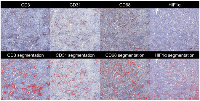
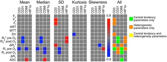
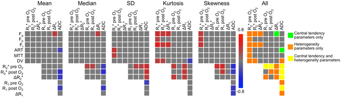
Tumour heterogeneity poses a significant challenge for treatment stratification. The goals of this study were to quantify heterogeneity in hepatocellular carcinoma (HCC) using multiparametric magnetic resonance imaging (mpMRI), and to report preliminary data correlating quantitative MRI parameters with advanced histopathology and gene expression in a patient subset. Thirty-two HCC patients with 39 HCC lesions underwent mpMRI including diffusion-weighted imaging (DWI), blood-oxygenation-level-dependent (BOLD), tissue-oxygenation-level-dependent (TOLD) and dynamic contrast-enhanced (DCE)-MRI. Histogram characteristics [central tendency (mean, median) and heterogeneity (standard deviation, kurtosis, skewness) MRI parameters] in HCC and liver parenchyma were compared using Wilcoxon signed-rank tests. Histogram data was correlated between MRI methods in all patients and with histopathology and gene expression in 14 patients. HCCs exhibited significantly higher intra-tissue heterogeneity vs. liver with all MRI methods (P < 0.030). Although central tendency parameters showed significant correlations between MRI methods and with each of histopathology and gene expression, heterogeneity parameters exhibited additional complementary correlations between BOLD and DCE-MRI and with histopathologic hypoxia marker HIF1α and gene expression of Wnt target GLUL, pharmacological target FGFR4, stemness markers EPCAM and KRT19 and immune checkpoint PDCD1. Histogram analysis combining central tendency and heterogeneity mpMRI features is promising for non-invasive HCC characterization on the imaging, histologic and genomics levels.
肿瘤异质性对治疗分层构成重大挑战。本研究旨在利用多参数磁共振成像(mpMRI)定量评估肝细胞癌(HCC)的异质性,并报告初步数据,这些数据将在患者亚组中关联定量 MRI 参数与高级组织病理学和基因表达。32 例 HCC 患者的 39 个 HCC 病变接受了 mpMRI 检查,包括弥散加权成像(DWI)、血氧水平依赖(BOLD)、组织氧水平依赖(TOLD)和动态对比增强(DCE)-MRI。使用 Wilcoxon 符号秩检验比较 HCC 和肝实质的直方图特征[中央趋势(均值、中位数)和异质性(标准差、峰度、偏度)MRI 参数]。在所有患者中,对直方图数据进行了所有 MRI 方法之间以及与 14 例患者的组织病理学和基因表达之间的相关性分析。与所有 MRI 方法相比,HCC 内部的组织内异质性明显更高(P<0.030)。尽管中央趋势参数与 MRI 方法之间以及与每种组织病理学和基因表达之间均显示出显著相关性,但异质性参数在 BOLD 和 DCE-MRI 之间以及与组织学缺氧标志物 HIF1α和 Wnt 靶基因 GLUL、药理靶基因 FGFR4、干细胞标志物 EPCAM 和 KRT19 以及免疫检查点 PDCD1 的基因表达之间显示出额外的补充相关性。联合中央趋势和异质性 mpMRI 特征的直方图分析有望在影像学、组织学和基因组学水平上实现非侵入性 HCC 特征描述。