Möllerherm Helene, Branitzki-Heinemann Katja, Brogden Graham, Elamin Ayssar A, Oehlmann Wulf, Fuhrmann Herbert, Singh Mahavir, Naim Hassan Y, von Köckritz-Blickwede Maren
Department of Physiological Chemistry, University for Veterinary Medicine Hannover, Hanover, Germany.
LIONEX Diagnostics & Therapeutics, Braunschweig, Germany.
Front Immunol. 2017 May 11;8:541. doi: 10.3389/fimmu.2017.00541. eCollection 2017.
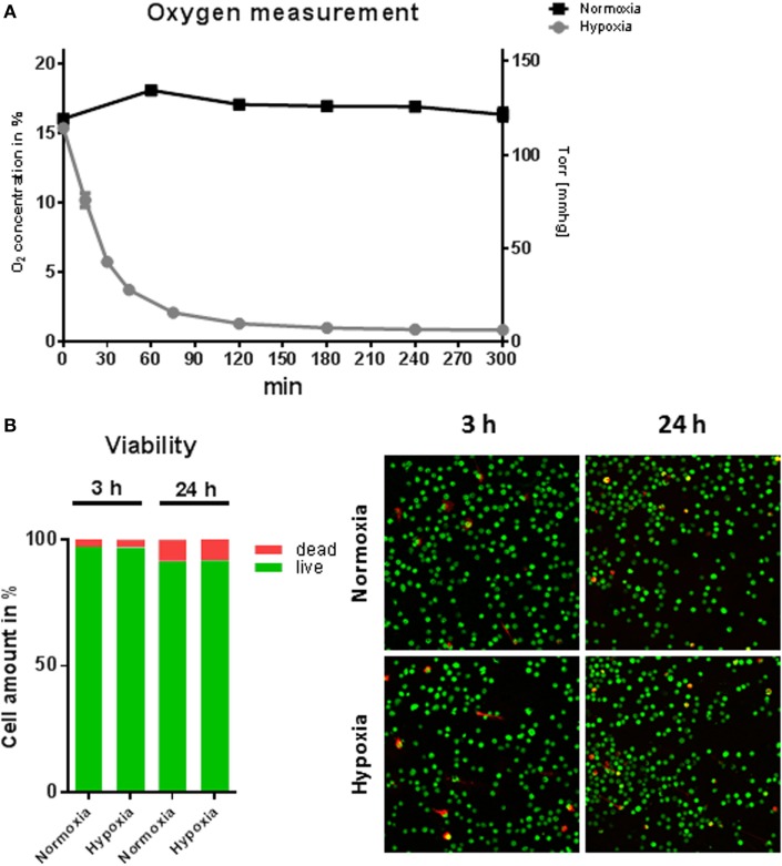
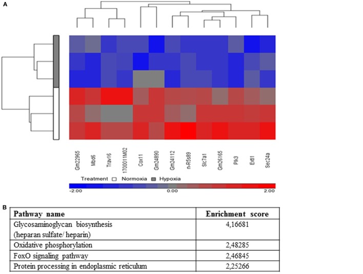
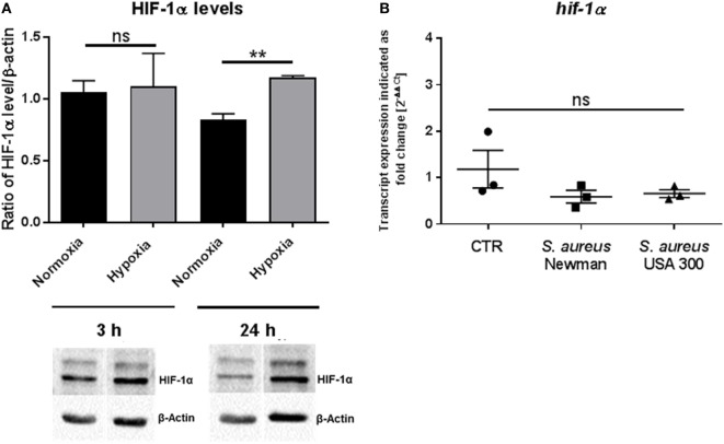
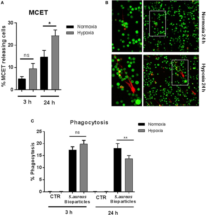
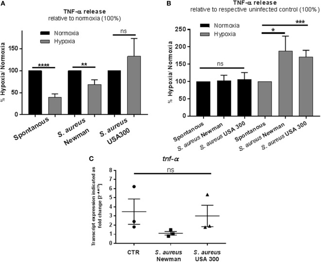
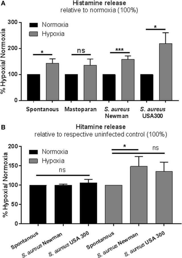
To study the antimicrobial function of immune cells , cells are commonly cultivated under atmospheric oxygen concentrations (20-21%; normoxia), although the physiological oxygen conditions are significantly lower in most tissues. Especially during an acute infection, oxygen concentration locally decreases to hypoxic levels around or below 1%. The goal of this study was to investigate the effect of hypoxia on the activity of mast cells (MCs). MCs were cultivated for 3 or 24 h at 1% O in a hypoxia glove box and co-incubated with heat-inactivated . When incubating the cells for 24 h under hypoxia, the transcriptional regulator hypoxia-inducible factor 1α (HIF-1α) was stabilized and resulted in increased extracellular trap formation and decreased phagocytosis. Interestingly, while phagocytosis of fluorescent bioparticles as well as the release of extracellular traps remained unaffected at 3 h hypoxia, the secretion of the prestored mediator histamine was increased under hypoxia alone. In contrast, the release of TNF-α was generally reduced at 3 h hypoxia. Microarray transcriptome analysis revealed 13 genes that were significantly downregulated in MCs comparing 3 h hypoxia versus normoxia. One interesting candidate is , a member of the pre-budding complex of coat protein complex II (COPII), which is responsible for the anterograde transport of proteins from the ER to the Golgi apparatus. These data lead to the suggestion that synthesized proteins including crucial factors, which are involved in the response to an acute infection like TNF-α, may eventually be retained in the ER under hypoxia. Importantly, the expression of HIF-1α was not altered at 3 h. Thus, our data exhibit a HIF-1α-independent reaction of MCs to short-term hypoxia. We hypothesize that MCs respond to short-term low oxygen levels in a HIF-1α-independent manner by downregulating the release of proinflammatory cytokines like TNF-α, thereby avoiding uncontrolled degranulation, which could lead to excessive inflammation and severe tissue damage.
为了研究免疫细胞的抗菌功能,尽管大多数组织中的生理氧条件明显较低,但细胞通常在大气氧浓度(20 - 21%;常氧)下培养。特别是在急性感染期间,局部氧浓度会降至1%左右或以下的缺氧水平。本研究的目的是调查缺氧对肥大细胞(MCs)活性的影响。肥大细胞在缺氧手套箱中于1% O₂条件下培养3小时或24小时,并与热灭活的……共同孵育。当在缺氧条件下将细胞孵育24小时时,转录调节因子缺氧诱导因子1α(HIF - 1α)会稳定下来,导致细胞外陷阱形成增加和吞噬作用降低。有趣的是,虽然在缺氧3小时时荧光生物颗粒的吞噬作用以及细胞外陷阱的释放未受影响,但仅在缺氧条件下预先储存的介质组胺的分泌增加。相比之下,在缺氧3小时时TNF - α的释放通常会减少。微阵列转录组分析显示,与常氧相比,在缺氧3小时的MCs中有13个基因显著下调。一个有趣的候选基因是……,它是COPII(II型被膜小泡蛋白复合物)预出芽复合体的成员,负责蛋白质从内质网到高尔基体的顺向转运。这些数据表明,包括参与急性感染反应如TNF - α等关键因子在内的合成蛋白质,在缺氧条件下最终可能会滞留在内质网中。重要的是,HIF - 1α的表达在3小时时未改变。因此,我们的数据显示MCs对短期缺氧有不依赖HIF - 1α的反应。我们假设MCs以不依赖HIF - 1α的方式对短期低氧水平作出反应,通过下调TNF - α等促炎细胞因子的释放,从而避免可能导致过度炎症和严重组织损伤的不受控制的脱颗粒。